Ukjent sin avatar

Slump Test: Sensory Responses in Asymptomatic Subjects

Om slump test for å finne ut av nevropatisk smerte eller betennelse.

http://www.ncbi.nlm.nih.gov/pmc/articles/PMC2565641/

«While non-neural structures such as subcutaneous connective tissues, skin, blood vessels, and fascia may also be placed under increasing loads during neural tissue testing, Coppieters et al demonstrated that successive stages of the Slump Test did not alter the perception of experimentally induced muscle pain (i.e., non-neural pain). This finding provides some validation for use of the Slump Test in the examination of neural structures.»

«The procedure was divided into four stages:

  • SS—the subject was asked to put her hands behind her back, to slump at the mid- and lower back, and to tuck her chin into the chest, while the examiner placed his hand at the cervicothoracic junction to monitor cervical position (Figure 1.2);
  • KE—while maintaining the above position, the subject was asked to extend the left knee until full extension was reached (Figure 1.3);
  • AD—the subject was then asked to dorsiflex the left ankle (Figure 1.4);
  • CE—the subject was asked to maintain the lower limb position while the examiner removed his hand from the cervicothoracic junction and the subject extended the neck (Figure 1.5).
  • The subject was then asked to assume a comfortable sitting position and any residual responses were recorded (Figure 1.6). The examiner practised the test procedure several times on one subject to increase the consistency in test application prior to data collection.»

«All responses were located in the back or neck for the 29.8% of subjects who experienced a sensory response during SS, but for the subsequent three stages of the test, the vast majority of responses were located in the thigh, knee, or calf (Table 1)»

«A positive Slump Test implicates neural tissue as the source of symptoms. As found in this study, responses other than the presenting symptoms may simply be an artefact of the test, typical of the normal response in asymptomatic subjects; such responses must not be assumed to indicate a positive Slump Test. Bilateral comparison is advocated in interpreting this test.»

Ukjent sin avatar

The effects of neurodynamic mobilization on fluid dispersion within the tibial nerve at the ankle: an unembalmed cadaveric study

Om hva nevropati og skader på nerver gjør, og hvordan neurodynamiske øvelser øker blodsirkulasjon internt i nerven. Nevner også hvordan skader, lav blodsirkulasjon og betennelser skaper sammensmeltninger i bindevevet mellom nerver og omliggende vev (muskler, skjelett, bindevev) som gjør at nervene ikke glir og dermed kan gi oss begrenset bevegelighet.

http://www.ncbi.nlm.nih.gov/pmc/articles/PMC3172954/

«These disorders include compression syndromes or other neuromuscular conditions that may be accompanied by neuropathic pain. Damaged nerves exhibit predictable pathophysiological responses including impaired nerve mobility, increased mechanosensitivity, impaired nerve conduction, nerve tissue ischemia, axonal transport inhibition, and intraneural edema.

The following popper user interface control may not be accessible. Tab to the next button to revert the control to an accessible version.  Impaired nerve mobility and increased mechanosensitivity provide the basis for existing studies of neurodynamic techniques. »

«Impaired nerve mobility and mechanosensitivity can be clinically assessed by measuring changes in joint range of motion, pain reproduction, or change of symptoms following neurodynamic mobilization.

«Intraneural edema is a common response to nerve injury, and is intimately involved in the proliferation of damage to nerve structure and function.

The following popper user interface control may not be accessible. Tab to the next button to revert the control to an accessible version. Edema is found in peripheral nerves that have been subject to trauma such as compression,excessive tension events, or vibration.  Even mild injury may result in epineurial edema,  but compression that is prolonged or of great magnitude leads to a breach of the diffusion barriers created by both the perineurium and microvasculature, resulting in endoneurial edema. The absence of lymphatic vessels in the endoneurium limits drainage of this edema, thereby creating a ‘mini-compartment syndrome’ within the nerve. »

«This ‘mini-compartment syndrome’, due to the increase in endoneurial pressure, subsequently leads to fibrosis and adhesions, impairing intrafascicular gliding. This loss of intrafascicular gliding creates an internal stretch lesion (Fig. 1). »

«The results showed significant mobilization effects in that there was increased fluid dispersion within the tibial nerve after the intervention. »

…de brukte bare kadavere i denne studien.
«The results showed significant mobilization effects in that there was increased fluid dispersion within the tibial nerve after the intervention. Because the tibial nerve was dissected free of all adjacent tissue and eliminated any external interfaces, the response to the mobilization appeared to be due to intraneural mechanics.»

Bevegelsene «pumper» internt i nerven og øker blodgjennomstreømning.
«During the mobilization technique, the tibial nerve alternately elongated and shortened which may have provided a temporary increase in intraneural pressure followed by a period of relaxation. Although speculative, it appears that this repetitive or ‘pumping’ action may have created a flushing of the dye and an alteration of the intraneural pressure as the intraneural fluid was dispersed.»

«In the early stages of stretch injury or compression, the ability to prevent or at least reduce edema may prevent or slow the inhibition of blood flow, thus preventing the sequelae leading to impaired axonal transport, demyelination, loss of elasticity due to fibrosis or adhesions, and ultimately to alteration in nerve structure and function. «

Ukjent sin avatar

Molecular profiling reveals synaptic release machinery in Merkel cells

Ett par studier om Merkel celler som reagerer på vibrasjon med lav frekvens (5-30 Hz) som sier mange ting om deres funksjon. Den sier bl.a. at de utløser glutamate, som er et nevropeptid relatert til smertetilstander. Men den utløser også andre nevrotransmittere som nedregulerer andre sanse-nevroner. Pluss at den innerverer eptielceller.
http://m.pnas.org/content/101/40/14503.full

«These data indicate that Merkel cells are poised to release glutamate and neuropeptides.»

«Because the question of whether Merkel cells are sensory cells is unresolved, other functions have been proposed. For example, Merkel cells may play a passive role in touch by efficiently transmitting force to mechanosensitive afferents (12). Alternatively, they may release neuromodulators to regulate the sensitivity of mechanoreceptive neurons (20). Merkel cells have also been proposed to influence the development or innervation of epithelia (ref. 21, but see ref. 22).»

«Together, these data demonstrate that Merkel cells are excitable cells and designate glutamate and CCK8 as candidate neurotransmitters at synapses between Merkel cells and sensory afferents in vivo. Our conclusion that Merkel cells function as excitable cells is strengthened by the abundance of neuronal transcription factors that we found to be enriched in Merkel cells (Table 1).»

«How might neurotransmitter release be stimulated from Merkel cells? Merkel cells may be mechanoreceptive cells that are directly activated by touch. Alternatively, Merkel cells may receive input from active afferent terminals. The latter conjecture is bolstered by reports of reciprocal connections in Merkel cell-neurite complexes (11).»

Ukjent sin avatar

Neuromuscular strain as a contributor to cognitive and other symptoms in chronic fatigue syndrome: hypothesis and conceptual model

Viktig studie om nervesystemet og bindevevets «adhesions» som bidragsyter til smerte.

http://www.frontiersin.org/Integrative_Physiology/10.3389/fphys.2013.00115/full

«Work by Brieg, Sunderland, and others has emphasized the ability of the nervous system to undergo accommodative changes in length in response to the range of limb and trunk movements carried out during daily activity. If that ability to elongate is impaired—due to movement restrictions in tissues adjacent to nerves, or due to swelling or adhesions within the nerve itself—the result is an increase in mechanical tension within the nerve. »

«This adverse neural tension, also termed neurodynamic dysfunction, is thought to contribute to pain and other symptoms through a variety of mechanisms. These include mechanical sensitization and altered nociceptive signaling, altered proprioception, adverse patterns of muscle recruitment and force of muscle contraction, reduced intra-neural blood flow, and release of inflammatory neuropeptides. »

«In our clinical work, we have found that neuromuscular restrictions are common in CFS, and that many symptoms of CFS can be reproduced by selectively adding neuromuscular strain during the examination.»

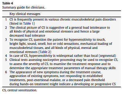
«As defined by Yunus, central sensitivity is “clinically and physiologically characterized by hyperalgesia (excessive sensitivity to a normally painful stimulus, e.g., pressure), allodynia (painful sensation to a normally non-painful stimulus, e.g., touch and massage), expansion of the receptive field (pain beyond the area of peripheral nerve supply), prolonged electrophysiological discharge, and an after-stimulus unpleasant quality of pain (e.g., burning, throbbing, numbness)” (Yunus, 2008).»

«These symptoms might be mediated by amplified central sensitivity, but peripheral factors, which have been described in FM and irritable bowel syndrome (IBS), may also play a role (e.g., Price et al., 2009; Staud et al., 2009). »

«Staud has shown that local anesthetic injection into trapezius muscle tender points results in lower levels of thermal hyperalgesia in the forearm, consistent with peripheral nociceptive input as a contributor to central sensitization (Staud et al., 2009).»

«The interaction of nerve mechanics and function has been termed neurodynamics. As an example of the principles of neurodynamics, the median nerve elongates approximately 20% as the upper extremity moves from a position of full wrist and elbow flexion to one of full wrist and elbow extension (Butler, 1991). »

» If that ability to elongate is impaired—due to movement restrictions in tissues adjacent to the median nerve and its branches, or due to swelling or adhesions within the median nerve itself—the result is an increase in mechanical tension within the nerve. This adverse neural tension, also termed neurodynamic dysfunction, is thought to contribute to pain and other symptoms through mechanical sensitization and altered nociceptive signaling, altered proprioception, adverse patterns of muscle recruitment and force of muscle contraction, reduced intra-neural blood flow, and release of inflammatory neuropeptides (Lindquist et al., 1973; Kornberg and McCarthy, 1992;Shacklock, 1995; Slater and Wright, 1995; Balster and Jull, 1997; Van der Heide et al., 2001; Kobayashi et al., 2003; Orlin et al., 2005).»

«It is now well-established that manual stretch of nerves is capable of evoking increased sweating and alterations of blood flow in peripheral tissues, providing evidence of electrophysiologic activity in sympathetic nerve fibers (Lindquist et al., 1973; Kornberg and McCarthy, 1992; Slater and Wright, 1995; Orlin et al., 2005). Conversely, treatment of areas of adverse neural tension (for example in carpal tunnel syndrome, cervico-brachial pain, and osteoarthritis) leads to improved functional outcomes (Rozmaryn et al., 1998; Deyle et al., 2000; Tal-Akabi and Rushton, 2000;Akalin et al., 2002; Allison et al., 2002).»

«The most notable examples of these provocation maneuvers are ankle dorsiflexion, the passive straight leg raise test, the upper limb tension (or neurodynamic) tests, and the seated slump test (Butler, 1991,2000). Test-retest reliability is good for straight leg raise, slump testing, and upper limb neurodynamic testing. (Coppieters et al., 2001;Herrington et al., 2008

«Because it is not possible to differentiate completely between adverse neural tension and strain in muscles, fascia, and other soft tissues, we will use the more general term “neuromuscular strain” in this paper. »

«As shown on the left in Figure 1, neuromuscular strains and movement restrictions can develop as a result injuries and activities of daily life (for example, due to soft tissue and peri-neural adhesions around scars, contusions and fractures that reduce range of motion, anatomic abnormalities like scoliosis and kyphosis, overuse injuries, and others).»

«If the neuromuscular strains were not treated, and if the individual adapted to the increased symptom burden with decreased activity, then neural, soft tissue and muscular restrictions would be expected to worsen, leading to greater impairment and greater central sensitization. »

«In our clinical work, we have found that neuromuscular restrictions are common in CFS.»
«We have also noted that many symptoms of CFS can be reproduced by selectively adding neuromuscular strain during the examination (Rowe et al., 2013a,b). »

«Despite the elevation of the leg, which might have been expected to improve venous return to the heart and thereby improve blood flow to the brain, lightheadedness increased, as did visual blurring. Both individuals remained more fatigued than usual for 12–24 h. Thus, supine neuromuscular strain provoked increased fatigue and cognitive disturbance, the two symptoms not adequately explained by the central sensitivity hypothesis thus far.»

«We have observed that open treatment of these movement restrictions using manual therapy is associated with clinical improvement (Rowe et al., 2013a,b).»

«The hypothesis can be tested by evaluating the whether the response to a given neuromuscular strain differs between CFS subjects and controls with regard to immediate and delayed (24-h) symptoms, and with regard to measures of central sensitivity, such as changes in heart rate variability, or changes in pain sensitivity as measured by pressure-pain thresholds. «

Ukjent sin avatar

Recognition of central sensitization in patients with musculoskeletal pain: Application of pain neurophysiology in manual therapy practice

Nevner det aller meste om sentral sensitering og hvordan de gjenkjennes i klinisk praksis.

http://www.thblack.com/links/RSD/ManTher2010_15_135_CSinMusculoskelPain-PainNeurophys.pdf

«Even with acute pain the nervous system undergoes some changes. When tissue is damaged and pain persists for a few days with adaptation of unimodal nociceptors, the responsiveness of polymodal nociceptive endings is enhanced by substances released from various sources (i.e. serotonin released by platelets) (Purves et al., 1997). This process is called primary hyperalgesia or peripheral sensitization of nociceptors, and represents a protective action by the human body in order to prevent further use of damaged structures and consequent further damage of the traumatized and surrounding tissues.»

«Secondary hyperalgesia refers to increased responsiveness of dorsal horn neurons localized in the spinal segments of the primary source of nociception.»

«Central sensitization is defined as an augmentation of responsiveness of central neurons to input from unimodal and polymodal receptors (Meyer et al., 1995). Central sensitization encompasses altered sensory processing in the brain (Staud et al., 2007), malfunctioning of descending anti-nociceptive mechanisms (Meeus et al., 2008), increased activity of pain facilitatory path- ways, temporal summation of second pain or wind-up (Meeus and Nijs, 2007; Staud et al., 2007), and long-term potentiation of neuronal synapses in the anterior cingulate cortex (Zhuo, 2007).»

«The presence of central sensitization in patients with musculoskeletal pain implies an increased complexity of the clinical picture (i.e. an increase in unrelated symptoms and hence a more difficult clinical reasoning process) (Nijs et al., 2009), as well as decreased odds for a favorable rehabilitation outcome (Jull et al., 2007).»

«Central sensitivity syndromes is a term first used by Yunus in 2000 to describe a group of overlapping conditions bound by a common pathophysiological mechanism of central sensitization (Yunus, 2007a).»

«Another example is chronic non-specific low back pain. Some studies provided evidence in support of the presence of central sensitization in patients with non-specific chronic low back pain (Giesecke et al., 2004; Schmidt-Wilcke et al., 2006), while others refute such an association (Hoffman et al., 2005; Julien et al., 2005). It is concluded that central sensitization is present in some cases of chronic non-specific low back pain, possibly representing one of the subgroups of this frequent musculoskeletal disorder (Wand and O’Connell, 2008).»

«The myofascial variety within the heterogeneous group of temporomandibular disorders is also characterized by central sensitization (Yunus, 2007a). Likewise, regional chronic pain conditions that present with tender and/or trigger points in the absence of structural pathology (frequently referred to as myofascial pain syndrome) should alert the manual therapist to the possibility that central sensitization is determining the clinical picture (Yunus, 2007a). However, to our knowledge available evidence in support of central sensitization in patients with myofascial pain syndrome is limited to chronic whiplash associated disorders, temporoman- dibular disorders and chronic non-specific low back pain.»

«Furthermore, various subgroups of headache, chronic tension-type headache (Langemark et al., 1993; Pielsticker et al., 2005) and migraine (Burnstein et al., 2000; Weissman-Fogel et al., 2003) can be viewed as central sensitivity syndromes. Finally, rheumatoid arthritis and osteoarthritis are examples of local musculoskeletal disorders possibly causing continuous activation of polymodal nociceptors that initiate or sustain central sensitization (Yunus, 2007a).»

table1

«Central sensitization entails much more than generalized hypersensitivity to pain: it is characterized by an increased responsiveness to a variety of stimuli including mechanical pressure (Desmeules et al., 2004), chemical substances (Morris et al., 1997), cold temperature (Kasch et al., 2005), heat temperature (Meeus et al., 2008), electrical stimuli (Banic et al., 2004; Desmeules et al., 2004), stress, emotions, and mental load. The clinical picture is suggestive of a general intolerance to all kinds of physical and emotional stressors and hence a large decreased load tolerance of the human body in general.»

table2

«An ongoing source of peripheral nociception is required before the process of peripheral sensitization can establish central sensitization (Nijs and Van Houdenhove, 2009). Tissue injury healing and focal pain recovery should occur as soon as possible to prevent development of central sensitization (Vierck, 2006).»

«One of the main characteristics of central sensitization in patients with musculoskeletal pain is a generalized rather than a localized decrease in their pressure pain threshold. Here, ‘generalized’ implies more than a segmental spreading of the symptom area, in that it means that the increased sensitivity is localized at sites segmentally unrelated to the primary source of nociception (e.g. the lower limbs in case of a whiplash trauma).»

«Lower pressure pain thresholds at symptomatic areas most often represent primary hyperalgesia due to sensitized polymodal noci- ceptors within injured musculoskeletal structures. By measuring pressure pain thresholds outside the area of primary nociception, widespread hyperalgesia or secondary hyperalgesia can be detec- ted.»

«In cases of secondary hyperalgesia, a reduced pressure pain threshold in the various tissues innervated by the same segment (or two neighboring segments) can be detected.»

«Findings of numerous areas of hyperalgesia at sites outside and remote from the symp- tomatic site, together with a non-segmental general decrease in pressure pain threshold, may imply a generalized hyperexcitability of central nociceptive pathways (Sterling et al., 2004).»

«Pressure algometry provides a reliable and valid measure of the pressure pain threshold (Vanderweeen et al., 1996; Farasyn and Meeusen, 2003). In the absence of a pressure algometer, manual palpation can be used. Even when a manual therapist is not sus- pecting central sensitization, the finding of generalized hypersen- sitivity to manual palpation during routine clinical examination should alert the clinician.»

«Like every other tissue in the human body, peripheral nerves and nervous tissues (including connective tissue) themselves can become hypersensitive to mechanical stimuli such as tension and pressure.»

«Besides the passive tests listed above (Table 3), altered sensory processing can be demonstrated during exercise. Pain thresholds increase during physical activity in healthy individuals and can stay augmented for up to 30 min post-exercise. This is the result of endogenous opioid release (Koltyn and Arbogast, 1998) and related activation of several (supra)spinal anti-nociceptive mechanisms such as the adrenergic and serotonergic pathways (Millan, 2002).»


«Stress (particularly when chronic) may well trigger lower pain thresholds. This was demonstrated by Suarez-Roca et al. (2008) who reported reduced GABA neurotransmission and consequent hyperalgesia in rats after repeated forced swimming stress.»

«A constant or decreased pain threshold during and following exercise suggests malfunc- tioning of these anti-nociceptive mechanisms (Whiteside et al., 2004) and hence central sensitization. An abnormal pain threshold response to exercise should be regarded as one of the many possible signs of central sensitization.»

table4


Ukjent sin avatar

Carbon dioxide influence on nitric oxide production in endothelial cells and astrocytes: Cellular mechanisms

Viktig studie som nevner hvordan CO2 forholder seg til NO og vasodilasjon. Nevner mekanismene bak eNOS og nNOS og hva som faktisk skjer i cellene. Denne studien er på celler, men beskriver mye av det som skjer in vivo og refererer til andre viktige studier.

http://www.ncbi.nlm.nih.gov/pmc/articles/PMC3073030/

«Cerebral vessels may regulate cerebral blood flow by responding to changes in carbon dioxide (CO2) through nitric oxide (NO) production. »

«NO levels in endothelial cells increased during hypercapnia by 36% in 8 hours and remained 25% above baseline. NO increase in astrocytes was 30% after 1 hour but returned to baseline at 8 hours. NLA blocked NO increase in endothelial cells under hypercapnia.»

«This study suggests that cerebral endothelial cells and astrocytes release NO under normocapnic conditions and NO production is increased during hypercapnia and decreased during hypocapnia independent of pH. Further, this demonstrates that endothelial cells may play a pivotal role in chemoregulation by modulating NOS activity.»

«Modulation of cerebral vascular tone in response to changes in the arterial partial pressure of carbon dioxide (pCO2) is defined as chemoregulation. In humans hypocapnia produces vasoconstriction resulting in decreased cerebral blood flow (CBF), whereas hypercapnia produces vasodilation and increased CBF (Lavi et al., 2003). »

«Using nitric oxide synthase (NOS) inhibitors, several in vivo studies have suggested that vasodilation in response to increased pCO2may be mediated by NO (Lavi et al., 2006). »

«Under hypercapnic conditions (pCO2 56.3±8.7 mmHg), NO concentration increased from baseline levels to a mean of 10±0.6×10⁻¹⁰M during the first 4 hours (Figure 1A). NO concentration peaked at 36% (10.2±0.5×10⁻¹⁰M) above baseline at 8 hours and stabilized 25% (9.4±0.5×10⁻¹⁰M) above baseline until completion of the experiment.»

«By plotting NO changes as a function of pCO2, we could disregard time as a variable in NO production (Figure 3) to establish that changes in NO levels correlate with changes in pCO2 (R=0.99).»

CO2 og NO

«Under hypercapnic conditions (pCO2 56.3±8.7 mmHg), human fetal astrocytes increased NO production by 30% over baseline values to a mean level of 2.5±1.2×10⁻¹⁰M in the first hour of hypercapnia (Figure 2). NO production then gradually decreased to control levels after 8 hours and remained at control levels for the remainder of the experiments.»

CO2 og NO i astrocytt

«The pH values were kept stable within a neutral gap under normocapnic (7.39±0.01), hypercapnic (7.36±0.02) and hypocapnic (7.40±0.01) conditions.»

» Stimulation of NOS in the endothelial cells is consistent with the NO-dependent vasodilation and increased CBF that occur in vivo during hypercapnia, as has been shown in rats (Iadecola, 1992) and in primates (Thompson et al., 1996). Decreased NO production by endothelial cells also correlates with the in vivo vasoconstrictive response to hypocapnia shown previously (Lavi et al., 2003;Thompson et al., 1996).»

«Thus, it is unlikely that eNOS is responsible for the early or fast phase response during chemoregulation in vivo. There are several explanations for this phenomenon. First, nitrite (NO2), being a storage pool of NO, can be reduced to NO under acidic and hypoxic conditions in vivo (Cosby et al., 2003). Under these conditions nitrite releases NO in the presence of deoxygenated hemoglobin in blood (Cosby et al., 2003;Nagababu et al., 2003) or neuroglobin (Burmester et al., 2000) in neurons acting as a nitrite reductase (Petersen et al., 2008). »

«The chemoregulatory response to CO2 changes in vivo is rapid, occurring on the order of milliseconds; our results did not demonstrate this component of the chemoregulatory response.»

«Cerebrovascular reactivity in response to CO2 is impaired in diabetic or hypertensive patients with endothelial dysfunction (Lavi et al., 2006), suggesting an important role for endothelial cells in modulating CBF response to CO2. »

«It has been reported that the ATP-sensitive K+ channels play a pivotal role in microvessel vasodilation of the cerebral cortex in response to decreased pH corresponding to mild hypercapnia and that a NOS inhibitor could not alter this vasodilation (Nakahata et al., 2003).»

Ukjent sin avatar

Alteration of substance P-mediated vasodilatation and sympathetic vasoconstriction in the rat knee joint by adjuvant-induced inflammation

Nevner at kronisk bentente ledd mister reaksjonsevnen på nerveimpulser og blodsirkulasjon. Kunne trengt å lese hele denne studien.

http://www.sciencedirect.com/science/article/pii/0304394094900027

«In control knees, nerve stimulation produced a frequency-dependent vasoconstruction»

«Chronically inflamed joints showed virtually no response to either nerve stimulation or SP application, suggesting a radical alteration in sympathetic and neuropeptidergic actions.»

Ukjent sin avatar

Nitric Oxide Inhibits Nociceptive Transmission by Differentially Regulating Glutamate and Glycine Release to Spinal Dorsal Horn Neurons

Mer om NO sin funksjon som både smertedempende og smerteøkende. Nevner alle faktorer som spiller inn i både nervesystem, ryggmarg og perifert. Viktig studie som også indirekte kan brukes i å forstå sammenhengen mellom hvordan CO2 og økning av eNOS (i blodkar) kan dempe nNOS (i nervesystemet).

http://www.jbc.org/content/286/38/33190.full

«Our findings suggest that spinal endogenous NO enhances inhibitory glycinergic input to dorsal horn neurons through sGC-cGMP-protein kinase G. Furthermore, NO reduces glutamate release from primary afferent terminals through S-nitrosylation of voltage-activated Ca2+ channels. Both of these actions probably contribute to inhibition of nociceptive transmission by NO at the spinal level.»

«Nitric oxide (NO) is freely diffusible across the cell membranes and is synthesized by the nitric-oxide synthase (NOS)2 from L-arginine and different cofactors.»

«The three NOS isoforms, including neuronal NOS (nNOS), endothelial NOS (eNOS), and inducible NOS (iNOS), have distinct structures and functions (1, 2). »

Refererer til Superoxid, den viktigste antioksidanten som CO2 beskytter. Og til peroxynitritt, den farligste oksidanten som CO2 også beskytter mot.

«The diverse effects of NO is commonly mediated through increased cGMP production upon activation of NO-sensitive soluble guanylyl cyclase (sGC), S-nitrosylation, tyrosine nitration, and the interaction with superoxide to form peroxynitrite (3,,5). »

Hvordan NO gir smerte, og at NO-hemmere demper smerte.:
«Some studies suggest that spinal NO is involved in the potentiation of nociception. For example, mechanical hypersensitivity induced by nerve injury or tissue inflammation is reduced by intrathecal administration of nNOS inhibitors and in nNOS-knock-out mice (6, 9, 10). »

Hvordan NO demper smerte:
«In contrast, other studies have shown that spinal NO plays a role in the inhibition of nociceptive processing. In this regard, intrathecal administration of L-arginine increases the mechanical nociceptive withdrawal threshold in rats (12). »

Mengden har mye å si for effekten, med lite er det smertedempende, men mye er det smerteøkende.
«For instance, it has been shown that intrathecal injection of low doses of L-arginine or NO donors reduce nociception, but L-arginine or NO donors at the high doses potentiates nociceptive responses to formalin injection or nerve injury (16, 17).»

Nevner at hvis én type NO er nedregulert blir en annen type oppregulert. Muligheten er da for at om eNOS (i blodårer) er lav pga dårlig blodsirkulasjon og lite CO2, kan nNOS (i nervesystemet) økes og dermed gi smerter. Ved å øke eNOS kan man da få mindre nNOS og dermed en indirekte smertedemping.
«In addition, it should be noted that in eNOS-, nNOS-, or iNOS-knock-out mice, an increase in the expression of other NOS isoforms in the spinal cord has been reported (18, 19). This compensatory up-regulation of other NOS subtypes in specific NOS isoform-knock-out mice further confounds the interpretation of the results.»

Om at NO både øker nocicepsjon og demper den:

«The discrepancy may result from the use of different pain models, the amount of NO produced locally, and the specific CNS sites involved. For example, NO-cGMP inhibits dorsal horn neuronal activity at the spinal level but excites spinal dorsal horn neurons at the supraspinal level (15, 39). Also, while low concentrations of NO inhibit NMDA receptor activity (40, 41), high concentrations of NO stimulate TRPV1 and TRPA1 receptors (42). »

«GABA and glycine are the two predominant inhibitory neurotransmitters in the spinal cord. Blocking GABAA or glycine receptors in the spinal cord induces pain hypersensitivity in rats (4344).»

«Of note, L-arginine and SNAP had similar effects on glycinergic sIPSCs and mIPSCs, suggesting that NO can potentiate glycine release from presynaptic terminals of interneurons in the spinal dorsal horn. Thus, our findings indicate that NO potentiates glycinergic input to spinal dorsal horn neurons to attenuate nociceptive transmission

«Glutamate is an excitatory neurotransmitter critically involved in nociceptive transmission in the spinal dorsal horn. In this study, we found that L-arginine and SNAP significantly inhibited the frequency of glutamatergic sEPSCs of lamina II neurons. L-Arginine and SNAP also consistently inhibited glutamatergic EPSCs evoked from primary afferents in most lamina II neurons.»

«Thus, it is unlikely that the inhibitory effect of NO on synaptic glutamate release to dorsal horn neurons is secondary to increased glycine release and stimulation of presynaptic glycine receptors (35). Our findings strongly suggest that NO attenuates synaptic glutamate release by inhibition of HVACCs at primary afferent terminals.»

«Another salient finding of our study is that NO potentiates glycinergic input and inhibits glutamatergic synaptic transmission in the spinal dorsal horn through distinct signaling pathways.»

«Therefore, our findings suggest that spinal NO primarily inhibits glutamate release through S-nitrosylation of HVACCs at primary afferent terminals.»

«In addition to its inhibitory effect on HVACCs shown in our study, NO can inhibit NMDA receptor currents in recombinant systems (6061) and in spinal lamina II neurons (41). Because both HVACCs and NMDA receptors are critically involved in nociceptive transmission, it seems difficult to explain the proposed pronociceptive role of NO at the spinal level. Of note, systemic use of NO or NO donors has been shown to reduce pain intensity caused by sickle cell crisis or diabetic neuropathy in patients (62, 63). We found in the present study that intrathecal administration of L-arginine or SNAP in rats significantly increased the nociceptive mechanical thresholds.»

«Hence, NMDA receptor activation could lead to increased NO production and release, and NO can diffuse to the presynaptic site to reduce glutamate release from primary afferent terminals. By reducing glutamatergic transmission, NO could serve as a feedback regulator to attenuate nociceptive transmission at the spinal level during painful conditions.»

«In summary, our study provides important new evidence that spinal NO potentiates inhibitory glycinergic input but reduces glutamatergic synaptic transmission between primary afferents and dorsal horn neurons through distinct signaling mechanisms. The opposing effects of NO on glutamatergic and glycinergic synaptic transmission could contribute to the antinociceptive effect of NO at the spinal level. This new information is important for our understanding of the synaptic actions underlying the antinociceptive effect of NO at the spinal level.»


Ukjent sin avatar

Pain and analgesia: The dual effect of nitric oxide in the nociceptive system.

Om at NO har en tosidig effekt som både smertedempende og smerteøkende.

http://www.ncbi.nlm.nih.gov/pubmed/21723953
«Nitric oxide is an important neurotransmitter involved in the nociceptive process and, in the dorsal horn of the spinal cord, it contributes to the development of central sensitization. On the other hand, experimental data have also demonstrated that NO inhibits nociception in the peripheral and also in the central nervous system. »

«In addition, it has been shown that nitric oxide mediates the analgesic effect of opioids and other analgesic substances. «