Immediate effects of breathing re-education on respiratory function and range of motion in chronic neck pain.

Å lære seg å bruke riktige pustemuskler gir mindre muskelspenninger og bedre bevegelighet i nakken. Om diafragma, den viktigste pustemuskelen, er svak eller på en eller annen måte ikke blir brukt nok, vil nakkemusker ta over store deler av pustefunksjonen. Dette kan være grunnlag til mange plager i nakken.

I denne studien gjorde 36 mennesker 30 minutter pustetrening. Smertenivåer og muskelspenninger ble redusert, og bevegelse i brystkassen og i nakken ble økt. 

Med enkle øvelser kan man få store resultater. Kun 30 minutter er nok! Om man gjør øvelser hver dag og diafragma blir sterke så trengs det mye mindre tid også.

http://www.ncbi.nlm.nih.gov/pubmed/25141528/

CONCLUSION:

Breathing re-education can change breathing patterns and increase chest expansion. This change leads to an improvement in CROM Positive consequences may result from the improvement in diaphragm contraction or reduced activity of accessory muscles.

High reliability of measure of diaphragmatic mobility by radiographic method in healthy individuals.

Nevner bevegelsen i diafragma under en spirometri test (innpust og utpust) og at noen har observert 9cm bevegelighet.

http://www.ncbi.nlm.nih.gov/pubmed/23778774

http://www.scielo.br/scielo.php?script=sci_arttext&pid=S1413-35552013000200128&lng=en&nrm=iso&tlng=en

There were large ranges of variation (in mm) in the obtained minimum and maximum values, and such variations were also reported in other studies8,6,24. Simon et al.13 observed a diaphragmatic value range from 0 to 85 mm, Houston et al.25 observed a range from 23 to 97 mm, Kantarci et al.27 observed a range from 25 to 84 mm and Boussuges et al.8 observed a range from 36 to 92 mm.


Måle Vitalkapasitet (lungevolum) med ballong

Denne videoen beskriver hvordan vital kapasitet måles enkelt med en ballong. En greit teknikk å bruke for å sjekke din fremgang med diafragma øvelsene i Verkstedet Breathing System.

Den nevner også hvordan man regner ut kroppens overflateareal, Body Surface Area: BSA = roten av ( (høyde (cm)*vekt (kg)) / 3600)

For så å kunne beregnes hva en vital kapsitet burde være (svaret vises i kubikkcentimeter, cm3):

Menn: BSA * 2500

Kvinner: BSA *  2000

Den virkelige vitalkapsiteten måles ved å blåse i en ballong (som er strekt ut først for å gjøre den mest mulig rund) 3 ganger, og så forholde seg til det største volumet.

Ballong diameteren viser lungevolum i denne grafen:

lungevolum ballong test

Man regner med at alt over 80% av forventet vitalkapsitet er normalt.

THE VALUE OF BLOWING UP A BALLOON

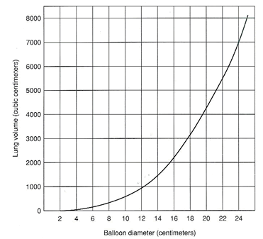
Dette er en veldig viktig artikkel for å forstå diafragmas rolle i både pust og bevegelse, og ifh smertetilstander i ryggraden. Nevner en lovende teknikk for å styrke diafragma og støttemuskulatur hvor man blåser opp en ballong og strammer kjernemuskulaturen. Nevner Zone of Apposition (ZOA) som beskriver diafragmas bevegelsesmuligheter. Ved lav ZOA har diafrgma lite bevegelse. Vi ønsker å øke ZOA. Denne øvelsen er konstruert basert på fysioterapeutisk prinsipper, men i Verkstedet Breathing System har vi øvelser som er gir samme resultater på diafragma, men bygget på lang og erfaringsbasert tradisjon fra tibetansk buddhisme.

Nevner også hvordan mage-pust minker bevegelsen i diafragma.

http://www.ncbi.nlm.nih.gov/pmc/articles/PMC2971640/

Suboptimal breathing patterns and impairments of posture and trunk stability are often associated with musculoskeletal complaints such as low back pain. A therapeutic exercise that promotes optimal posture (diaphragm and lumbar spine position), and neuromuscular control of the deep abdominals, diaphragm, and pelvic floor (lumbar-pelvic stabilization) is desirable for utilization with patients who demonstrate suboptimal respiration and posture. This clinical suggestion presents a therapeutic exercise called the 90/90 bridge with ball and balloon. This exercise was designed to optimize breathing and enhance both posture and stability in order to improve function and/or decrease pain. Research and theory related to the technique are also discussed.

Many muscles used for postural control/stabilization and for respiration are the same, for example: the diaphragm, transversus abdominis, and muscles comprising the pelvic floor.16 Maintaining optimal posture/stability and respiration is important and is even more challenging during exercise. Exercise increases respiratory demand (e.g. running) and limb movements (e.g. arms moving while standing still) increase postural demands for stabilization.3,7

Many factors are potentially involved with suboptimal respiration and suboptimal (faulty) posture and may be associated with musculoskeletal complaints such as low back pain, and/or sacroiliac joint pain.8 (Table 1)

Suboptimal Respiration and Posture
Decreased/suboptimal Zone of Apposition of diaphragm
Decreased exercise tolerance
Decreased intra-abdominal pressure
Shortness of Breath/Dyspnea
Decreased respiratory efficiency
Decreased expansion of lower rib cage/chest
Decreased appositional diaphragm force
Decreased length of diaphragm (short)
Decreased transdiaphragm pressure
Increased use of accessory muscles of respiration
Poor neuromuscular control of core muscles
Increased lumbar lordosis
Increased anterior pelvic tilt
Increased hamstring length
Increased abdominal length
Rib elevation/external rotation
Sternum elevation
Increased activity of paraspinals
Increased lumbar-pelvic instability
Low back pain
Sacroiliac Joint pain
Thoracic Outlet Syndrome
Headaches
Asthma

One of the most critical factors, often overlooked by physical therapists, is maintaining an optimal zone of apposition of the diaphragm.3,911 The zone of apposition (ZOA) is the area of the diaphragm encompassing the cylindrical portion (the part of the muscle shaped like a dome/umbrella) which corresponds to the portion directly apposed to the inner aspect of the lower rib cage.12 The ZOA is important because it is controlled by the abdominal muscles and directs diaphragmatic tension. When the ZOA is decreased or suboptimal, there are several potential negative consequences. (Table 1) Two examples include:

  1. Inefficient respiration (less air in and out) because the transdiaphragmatic pressure is reduced.11 The smaller the ZOA, there will be less inspiratory action of the diaphragm on the rib cage.11
  2. Diminished activation of the transversus abdominis which is important for both respiration and lumbar stabilization.11,13

The incidence of LBP has been documented to be as high as 30% in the athletic population, and in many cases pain may persist for years.15 Low back pain is frequently correlated with faulty posture such as an excessive lumbar lordosis.1618 Excessive lumbar lordosis may be associated with over lengthened and weak abdominal musculature.1820 Poor neuromuscular control of core muscles (transversus abdominis, internal oblique, pelvic floor and diaphragm) has been described in individuals with SIJ pain21 and in individuals with lumbar segmental instability, potentially adversely affecting respiration.22

Richardson et al.27 describe coordination of the Transversus abdominis and the diaphragm in respiration during tasks in which stability is maintained by tonic activity of these muscles. During inspiration, the diaphragm contracts concentrically, whereas the transversus abdominis contracts eccentrically. The muscles function in reverse during exhalation with the diaphragm contracting eccentrically while the transversus abdominis contracts concentrically. Hodges et al. noted that during respiratory disease the coordinating function between the transversus abdominis and diaphragm was reduced.6 Thus, it is also possible that faulty posture such as over lengthened abdominals and excessive lordosis could reduce the coordination of the diaphragm and transversus abdominis during respiration and stabilization activities.

O’sullivan et al.21 studied subjects with LBP attributed to the sacroiliac joints and compared them to control subjects without pain. O’sullivan et al. compared respiratory rate and diaphragm and pelvic floor movement using real time ultrasound during a task that required load transfer through the lumbo-pelvic region (the active straight leg raise test). Subjects with pain had an increase in respiratory rate, descent of their pelvic floor and a decrease in diaphragm excursion as compared to the control subjects, who had normal respiratory rates, less pelvic floor descent, and optimal diaphragm excursion. While O’sullivan et al. concluded that an intervention program focused on integrating control of deep abdominal muscles with normal pelvic floor and diaphragm function may be effective in managing patients with LBP,21 they did not describe strategies or exercises to achieve this goal.21

While the role of the Transversus abdominis in lumbar stability is well documented, less well known is the role of the diaphragm in lumbar stability. While the primary function of the diaphragm is respiration, it also plays a role in spinal stability.3,28

The right hemidiaphragm attaches distally to the anterior portions of the first through third lumbar vertebrae (L1-3) and the left hemidiaphragm attaches distally on the first and second lumbar vertebrae (L1-2).29 This section of the diaphragm is referred to as the crura. Of interest is the asymmetrical attachment of the diaphragm with the left hemidiaphragm attaching to L1-2 and the right portion attaching to L1-3.

During the inhalation phase of ventilation, the dome of the diaphragm moves caudally like a piston creating a negative pressure in the thorax that forces air into the lungs. This action is normally accompanied by a rotation of the ribs outward (external rotation) largely in part due to the ZOA.12 (Figure 1) Apposition is a term that means multiple layers adjacent to each other.33 The normal force of pull on the sternal and costal portions of the diaphragm would produce an internal rotation of the ribs. The ZOA creates an external rotation of these ribs primarily because the pressure in the thoracic cavity prevents an inward motion. The crural portion of the diaphragm assists the caudal motion of the dome. It also pulls the anterior lumbar spine upward (cephalad and anterior). Additionally, the abdominal muscles and pelvic floor musculature are less active to allow visceral displacement due to the dome of the diaphragm dropping. With exhalation, this process is reversed. Abdominal muscle activity compresses the viscera in the abdominal cavity, the diaphragm is forced cephalad and the ribs internally rotate. As exhalation becomes forced as during exercise, abdominal activity (rectus abdominus, internal obliques, external obliques, and transversus abdominis) will be increased.3436

When the ZOA is optimized, the respiratory and postural roles of the diaphragm have maximal efficiency.37 In suboptimal positions (i.e. decreased ZOA), the diaphragm has a decreased ability to draw air into the thorax because of less caudal movement upon contraction and less effective tangential tension of the diaphragm on the ribs and therefore lower transdiaphragmatic pressure.38 This decreased ZOA is accompanied by decreased expansion of the rib cage, postural alterations, and a compensatory increase in abdominal expansion.12 (Figure 2)

One such adaptive breathing strategy would be to relax the abdominal musculature more than necessary on inspiration to allow for thoraco-abdominal expansion. This situation leads to decreased abdominal responsibility while breathing and can contribute to instability. This would reflect more upper chest breathing and less efficient diaphragm activity. If the body maintains this position and breathing strategy for an extended period of time, the diaphragm may adaptively shorten and the lungs may become hyperinflated.37,39,40 Hyperinflation may also contribute to over use of accessory muscles of respiration such as scalenes, sternocleidomastoid (SCM), pectorals, upper trapezius and paraspinals in an attempt to expand the upper rib cage.4144 Again, without an optimal dome shape/position of the diaphragm or an optimal ZOA the body compensates to get air in with accessory muscles since the more linear/flat/short diaphragm is less efficient for breathing.32

Instructions for Performance of the 90/90 Bridge with Ball and Balloon: 1. Lie on your back with your feet flat on a wall and knees and hips bent at a 90-degree angle. 2. Place a 4-6 inch ball between your knees. 3. Place your right arm above your head and a balloon in your left hand. 4. Inhale through your nose and as you exhale through your mouth, perform a pelvic tilt so that your tailbone is raised slightly off the mat. Keep low back flat on the mat. Do not press your feet into the wall, instead pull down with your heels. 5. You should feel the back of your thighs and inner thighs engage, keeping pressure on the ball. Maintain this position for the remainder of the exercise. 6. Now inhale through your nose and slowly blow out into the balloon. 7. Pause three seconds with your tongue positioned on the roof of your mouth to prevent airflow out of the balloon. 8. Without pinching the neck of the balloon and keeping your tongue on the roof of your mouth, inhale again through your nose. 9. Slowly blow out as you stabilize the balloon with your left hand. 10. Do not strain your neck or cheeks as you blow. 11. After the fourth breath in, pinch the balloon neck and remove it from your mouth. Let the air out of the balloon.12. Relax and repeat the sequence 4 more times. Copyright © Postural Restoration Institute™ 2009, used with permission

The patient/athlete is asked to hold the balloon with one hand and inhale through his/her nose with the tongue on the roof of the mouth (normal rest position) and then exhale through his/her mouth into the balloon. The inhalation, to about 75% of maximum, is typically 3-4 seconds in duration, and the complete exhalation is usually 5-8 seconds long followed by a 2-3 second pause. This slowed breathing is thought to further relax the neuromuscular system/parasympathetic nervous system and generally decrease resting muscle tone. Ideally the patient/athlete will be able to inhale again without pinching off the balloon with their teeth, lips, or fingertips. This requires maintenance of intra-abdominal pressure to allow inhalation through the nose without the air coming back out of the balloon and into the mouth.

When the exercise is performed by the patient/athlete with hamstring and gluteus maximus (glut max) activation (hip extensors) the pelvis moves into a relative posterior pelvic tilt and the ribs into relative depression and internal rotation. This pelvic and rib position helps to optimize abdominal length (decreases) and diaphragm length/ZOA (increases).

Clinical experience with the BBE includes utilization of the exercise for both female and male patients (more females than males), ages 5-89 with a wide variety of diagnoses including: low back pain, trochanteric bursitis, SIJ pain, asthma, COPD, acetabular labral tear, anterior knee pain, thoracic outlet syndrome (TOS) and sciatica.

om hvordan sittestilling påvirker kroppen

En gjennomgang av hvordan sitteposisjon og holdning påvirker kroppen.

http://www.dynamicchiropractic.ca/mpacms/dc_ca/article.php?id=56598

In order to assess the loads placed on a spine during various positions, Rohlmann, et al. (2011) looked at various seating positions.4 They found the implant force increased 48 percent for 15 degrees flexion and decreased 19 percent for 10 degrees extension of the trunk. Placing the hands on the thighs reduced the loads by 19 percent, on average, compared to having arms hanging at the sides.

Dreischarf, et al. (2010) also found that reduced spinal load during sitting can be achieved by supporting the upper body with the arms.5

A study by De Carvalho, et al. (2010) compared lumbar spine and pelvic posture between standing and sitting via radiologic investigation. Lumbar lordosis and sacral inclination decreased by 43 and 44 degrees, respectively.6 This shows that with respect to sitting posture, to goal should be to maintain or prevent a reduction of the lumbar lordosis.

One study found 40-percent higher cervical extensor activity in the slouched posture. More neutral sitting postures reduce the demand on the cervical extensor muscles.7 Education on maintaining a neutral sitting posture can offset the detrimental effects.

A study by Caneiro, et al. (2010) showed that slumped sitting was associated with greater head / neck flexion, and increased muscle activity of the cervical erector spinae.9 Adjustments to seat angle and lumbar roll can also significantly effect head and neck posture.

A study by Horton, et al. (2010) found that the degree of angulation of the backrest support of an office chair, plus the addition of a lumbar roll support, are the two most important seat factors that will benefit head and neck postural alignment.10

A study by Bullock, et al. (2005) looked at how sitting posture can affect range of motion and pain for those with shoulder impingement.11 An erect posture appeared to increase active shoulder flexion, although there was no difference in shoulder pain between an erect and slouched posture.

Finley, et al. (2003) found that an increased thoracic kyphosis from a slouched posture can significantly alter the kinematics of the scapula during humeral elevation.12

And Kebaetse, et al. (1999) found that a slouched posture is associated with a 16.2 percent reduction in arm horizontal muscle force.13

A recent study by Dunk, et al. (2009), out of the University of Waterloo, evaluated whether the intervertebral joints of the lumbosacral spine approach their end ranges of motion in a seated posture.15 In upright sitting, the L5-S1 intervertebral joint was flexed to more than 60 percent of its total range of motion. In a slouched posture, each of the lower three intervertebral joints approached their total flexion angles. This shows an increased loading of the passive tissues (time-dependent «creep»), which may contribute to low back pain from prolonged sitting.

A study by Reeve, et al. (2009) assessed the thickness of the TrA in various postural positions. Thickness was significantly greater in standing and erect sitting than in a slouched or sway-back standing position.16 The authors concluded that lumbopelvic neutral postures have a positive influence on spinal stability compared to equivalent poor postures.

A study by Claus, et al. (2009) looked at the effect of various postures on regional muscle activity.17 For the deep and superficial fibers of lumbar multifidus muscles, the least muscle activity occurred during a flat posture, which was similar to a slump posture. The most activity occurred in a short lordosis position; there was also more activity in the obliquus internus.

A study by Dolan, et al. (2006) provided evidence that a slouched posture of 5 minutes’ duration can increase reposition error.18 Proprioceptive control is known to be valuable in spinal stability. The fact that reposition error can occur within as little as 5 minutes of «slouched» posture suggests the importance of postural education in decreasing proprioceptive loss and injury.

Respiratory weakness in patients with chronic neck pain.

Studie som nevner at alle med kroniske nakkeplager også har svake pustemuskler, og at pusten kan bidra til å opprettholde smertene. Spesielt ved svak utpust (MEP – maximal expiratory pressure) er det sammenheng med nakkesmerter. Kunne trengt hele denne studien.

http://www.ncbi.nlm.nih.gov/pubmed/23199797

Neck muscle strength (r > 0.5), kinesiophobia (r < -0.3) and catastrophizing (r < -0.3) were significantly associated with maximal mouth pressures (P < 0.05), whereas MEP was additionally negatively correlated with neck pain and disability (r < -0.3, P < 0.05).

It can be concluded that patients with chronic neck pain present weakness of their respiratory muscles. This weakness seems to be a result of the impaired global and local muscle system of neck pain patients, and psychological states also appear to have an additional contribution. Clinicians are advised to consider the respiratory system of patients with chronic neck pain during their usual assessment and appropriately address their treatment.

Inspiratory muscle training reduces blood lactate concentration during volitional hyperpnoea

Med pustetrening blir melkesyrekonsentrasjonen lavere under trening. 2 studier her, første fra 2008 og den andre fra 2012.

Den første nevner at melkesyre synker med opptil 59% (25+34%)

http://www.ncbi.nlm.nih.gov/pubmed/18560878

Although reduced blood lactate concentrations ([lac(-)](B)) have been observed during whole-body exercise following inspiratory muscle training (IMT), it remains unknown whether the inspiratory muscles are the source of at least part of this reduction.

After 6 weeks, increases in [lac(-)](B) during volitional hyperpnoea were unchanged in the control group. Conversely, following IMT the increase in [lac(-)](B) during volitional hyperpnoea was reduced by 17 +/- 37% and 25 +/- 34% following 8 and 10 min, respectively (P < 0.05).

These findings suggest that the inspiratory muscles were the source of at least part of this reduction, and provide a possible explanation for some of the IMT-mediated reductions in [lac(-)](B), often observed during whole-body exercise.

Inspiratory muscle training abolishes the blood lactate increase associated with volitional hyperpnoea superimposed on exercise and accelerates lactate and oxygen uptake kinetics at the onset of exercise.

Den andre viser til en 15% laver melkesyrekonsentrasjon og at årsaken er pustemusklenes evne til å fjerne det.

http://www.ncbi.nlm.nih.gov/pubmed/21964908

Following the intervention, maximal inspiratory mouth pressure increased 19% in the IMT group only (P < 0.01). Following IMT only, the increase in [lac(-)](B) during volitional hyperpnoea was abolished (P < 0.05). In addition, the blood lactate (-28%) and phase II oxygen uptake (-31%) kinetics time constants at the onset of exercise and the MLSS [lac(-)](B) (-15%) were reduced (P < 0.05). We attribute these changes to an IMT-mediated increase in the oxidative and/or lactate transport capacity of the inspiratory muscles.

Inspiratory muscle training lowers the oxygen cost of voluntary hyperpnea

Nevner at innpustmuskel trening gir mindre oksygenbehov under trening og dermed mer utholdenhet. Innpustmuskler bruker opp mye av oksygenet kroppen trenger under trening så med svak pustefunksjon blir man fort sliten. Under maksimal trening krever pustemusklene 15% av oksygenet, men med pustetrening synker det til 8%. Den nevner at diafragma og pustemuskler blir sterkere og større. Den henviser også til studier som nevner at det gir mindre melkesyre. Noe av effekten kommer også av at man får en større reserve i lungene ved å øke inn- og utpust styrken.

http://jap.physiology.org/content/112/1/127.full

IMT significantly reduced the O2 cost of voluntary hyperpnea, which suggests that a reduction in the O2 requirement of the respiratory muscles following a period of IMT may facilitate increased O2 availability to the active muscles during exercise. These data suggest that IMT may reduce the O2cost of ventilation during exercise, providing an insight into mechanism(s) underpinning the reported improvements in whole body endurance performance; however, this awaits further investigation.

THE OXYGEN COST of breathing or energy requirement of the respiratory muscles are shown to increase relative to the level of ventilation (V̇E) and the work of breathing (Wb) (1, 8). During moderate-intensity exercise the respiratory musculature requires ∼3–6% of total oxygen consumption (V̇O2T), increasing to ∼10–15% at maximal exercise (1, 3).

Inspiratory muscle training (IMT) is an intervention that has been associated with improvements in whole body exercise performance (24, 31, 34), enhanced pulmonary oxygen uptake kinetics (5), reduced blood lactate concentrations (6, 24), diaphragmatic fatigue, and cardiovascular responsiveness (37).


The oxygen cost of voluntary hyperpnea (V̇O2RM) and V̇O2RM expressed as a percentage of total oxygen consumption (V̇O2T) graphed against V̇E at low (50% V̇O2 max), moderate (75% V̇O2 max), and high (100% V̇O2 max) exercise intensities for both IMT (A) and CON (B) groups, pre- and post-training (means ± SE).
•, Pre-IMT;
○, post-IMT;
▴, pre-CON;
Δ, post-CON.

To our knowledge this study is the first to investigate the influence of IMT on the oxygen cost of voluntary hyperpnea. The main findings of the present study are that the relationship between increasing ventilatory workloads and the O2 cost of voluntary hyperpnea is curvilinear in trained cyclists and that 6 wk of pressure threshold IMT significantly reduced the O2 cost of V̇E at high ventilatory workloads. Importantly, the finding that V̇O2RM is reduced at a V̇E above 50% V̇O2 max suggests that IMT may reduce the energy requirements of the respiratory musculature in maintaining a given V̇E.

The increase in energy expenditure as V̇E increases can be attributed to a variety of sources of respiratory muscle work, including the elastic recoil of the chest and lung wall, airway resistance (4,15), increased EELV (9), and high muscle shortening velocities (19, 23). It has been suggested that as tidal breathing approaches the maximal limits for inspiratory muscle pressure development and expiratory flow rates, energy expenditure may increase to overcome the additional respiratory muscle work (3). Conversely, if one or more of the additional sources of respiratory muscle work are reduced as a result of IMT, it is reasonable to suggest that the increase in the O2 cost maybe attenuated.

In the present study, following 6 wk of IMT, V̇O2RM was significantly reduced from pretraining values at submaximal and maximal levels of ventilation. The O2 cost of voluntary hyperpnea expressed as a percentage of V̇O2T was reduced by 1.5% at a V̇Ecorresponding to 75% V̇O2max following IMT. The greatest reduction in the O2 cost of voluntary hyperpnea was observed at V̇O2 max, where V̇O2RM was significantly reduced from 11% of V̇O2T to 8% V̇O2T following IMT.

Increased ventilatory demand was previously shown to elicit a sympathetically mediated metaboreflex (33), which increases heart rate and mean arterial pressure (MAP), reducing blood flow to the limb locomotor muscles during exercise (16) and potentially reducing whole body endurance performance (18). Furthermore, Witt et al. (37) showed that IMT attenuates this increase in HR and MAP, presumably by reducing or delaying the sympathetically mediated reflex.

The 22% increase in respiratory muscle strength shown in the present study is similar in magnitude to those previously reported using pressure-threshold IMT (11, 22, 30, 32, 37). Respiratory muscle structure has also been shown to change following IMT, with an increase in diaphragm thickness (11, 12) and hypertrophy of type II muscle fibers of the external intercostal muscles (27) being reported.

Aaron et al. (3) demonstrated that individuals who reached their reserve for expiratory flow and inspiratory muscle pressure development required 13–15% of V̇O2T compared with ∼10% of V̇O2T for non-flow-limited individuals. Thus, an increase in maximal expiratory flow rates or inspiratory pressure development would increase the ventilatory reserve, thereby increasing the maximal limits for ventilation.

Energy cost of breathing at depth: effect of respiratory muscle training

Om at å trene innpustmuskler gjør det lettere å puste normale og uanstrengt etterpå.

http://www.ncbi.nlm.nih.gov/pubmed/22908839

RRMT significantly reduced the energy cost of ventilation, measured as delta VO2/delta V(E) during ISEV, at a depth of 55 fsw. Whether this change was due to reduced work of breathing and/or increased efficiency of the respiratory muscles remains to be determined.