Ukjent sin avatar

Arterial Blood Gas Changes During Breath-holding From Functional Residual Capacity

Endelig en studie som viser direkte hva som skjer med CO2 når vi holder pusten etter utpust. Her kaller de det etter «functional residual capacity» (wikipedia), altså etter en normal og passiv utpust. Denne studien viser først og fremst hva som skjer i løpet av én enkelt pustehold. De nevner også at det tar 5-10 sekunder etter innpust igjen før CO2 nivet begynner å synke. Så i metablsk pust (RecoveryBreathing) hvor vi puster 10sek inn/ut og har 10 sek pause bør CO2 lett kunne stabiliseres på et høyere nivå enn normalt.

http://www.ncbi.nlm.nih.gov/pubmed/8874252

Hele Studien: http://journal.publications.chestnet.org/data/Journals/CHEST/21737/958.pdf

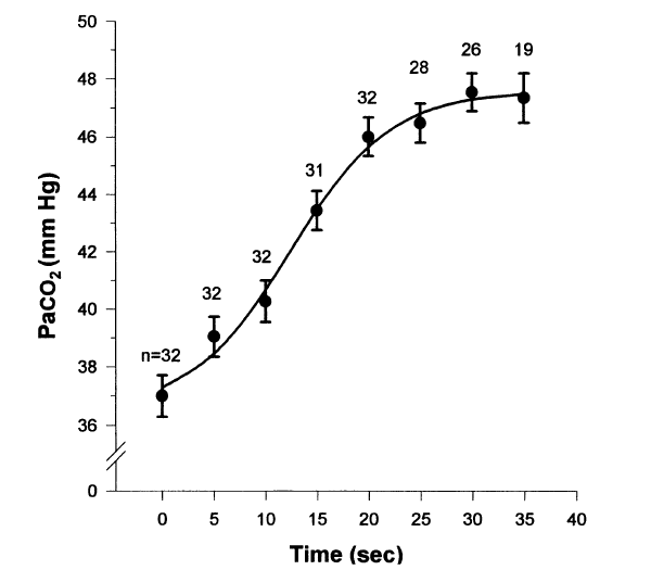
Breath-holding serves as a model for studying gas exchange during clinical situations in which cessation of ventilation occurs. We chose to examine the arterial blood gas changes that occurred during breath-holding, when breath-holding was initiated from functional residual capacity (FRC) while breathing room air. Eight normal subjects who had a radial artery catheter placed for another study were taught to breath-hold on command from FRC. FRC was determined using respiratory inductance plethysmography. Arterial blood gas specimens were obtained at 5-s intervals until the termination of breath-holding. The average breath-holding time (+/-SD) was 35 (+/-10 s). The PaO2, PaCO2, and pH values were plotted against time and individually fit to logistic equations for each subject. The arterial PaO2 fell by a mean of 50 mm Hg during the first 35 s of breath-holding under these conditions, while the arterial PCO2 rose by a mean of 10.2 mm Hg during the first 35 s and the pH fell by a mean of 0.07 in the first 35 s. The rapid decline in PaO2 is greater than that previously reported using different methods and should be considered in clinical situations in which there is an interruption of oxygenation and ventilation at FRC while breathing room air. The changes in PaCO2 and pH are similar to those previously reported in paralyzed apneic patients.

CO2 etter utpust

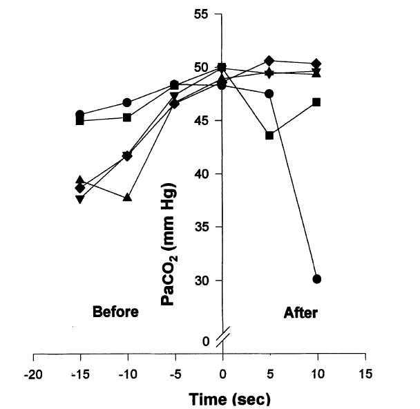
We demonstrated during 5 breath-holding runs in which additional arterial specimens were obtained at 5 and 10 s after breath-hold (Fig 6) that the elevated arterial PCO2 did not begin to fall until at least 5 s after breaking from the breath-hold in 1 run and greater than 10 s in 3 other runs. This implies that the removal of the remaining arterial PCO2 by the lungs took longer than 5 s before recirculation from pulmonary capillary blood could lower the arterial PCO2 in the radial artery. The second less significant factor that explains the persistent elevation of arterial PCO2 is the concentrating effect caused by the decreasing LV. The concentrating effect occurs with breath-holding, as more oxygen is removed from the lungs than carbon dioxide is added. As carbon dioxide production continues to occur, the capillary

CO2 fall etter innpust

Ukjent sin avatar

CO2 sin relasjon til pH

Tabell som viser sammenhengen mellom CO2 og pH. CO2 mellom 35-45 er normalt, over eller under dette kaller man det alkalose eller acidose. Med Metabolsk Pust (RecoveryBreathing.com) ønsker vi å få CO2 opp mot 40-50 mmHg. Legg merke til at også HCO3- økes når CO2 økes i blod. Bikarbonat kalles det og er det stoffet som er i Natron.

Klikk for å få tilgang til Oxygenation%20and%20oxygen%20therapy.pdf

TABLE IV

PaCO2 (mm Hg)

pH

HCO3-

15

7.61-7.74

15.3-20.5

20

7.55-7.66

17.7-22.8

30

7.45-7.53

21.0-25.6

40

7.38-7.45

22.8-26.8

50

7.31-7.36

24.1-27.5

60

7.24-7.29

25.1-27.9

70

7.19-7.23

25.7-28.5

80

7.14-7.18

26.2-28.9

90

7.13-7.09

Tabell som viser hvordan O2 og CO2 synker når man kommer opp i høyden.

TABLE V. Gas Pressures at Various Altitudes*

LOCATION

ALT.

PB

FIO2

PIO2

PaCo2

PAO2

PaO2

Sea Level

0

760

.21

150

40

102

95

Cleveland

500

747

.21

147

40

99

92

Denver

5280

640

.21

125

34

84

77

*Pikes’s Peak

14114

450

.21

85

30

62

55

*Mt. Everest

29028

253

.21

43

7.5

35

28

*All pressures in mm Hg; Pike’s Peak and Mt. Everest data from summits

Ukjent sin avatar

Elevated CO2 Levels Cause Mitochondrial Dysfunction and Impair Cell Proliferation

Økt CO2 i cellene gjør at celledeling går tregere. Dette kan fungere også bland kreftceller slik at de deler seg tregere og utvikler seg saktere. De kaller det mitokondrial dysfunksjon i overskriften, men dette er snakk om langvarig hyperkapni opp mot 100 mmHg, langt mer enn hva er mulig å få til med pusteøvelser (50-60 mmHg i korte perioder). Evnen til å kunne øke CO2 i korte perioder vil dermed kunne senke hastigheten på aldringsprosessen

Klikk for å få tilgang til 37067.full.pdf

As shown in Fig. 1, we found a decreased rate of proliferation (assessed as num- ber of cells (Fig. 1, A and B) and BrdU incorporation (Fig. 1, C and D)), which became significant after 3 days of exposure to high levels of CO2. Proliferation was decreased in a dose-depen- dent manner. It is also important to stress that the decreased proliferation was independent of extracellular pH.

Decreased Cell Proliferation during Exposure to High CO2 Levels Is Not Due to Increased Cell Death or Cell Cycle Arrest.

We found that exposure to high CO2 did not result in increased cell death.

We also did not find differences in the distribution of cell cycle phases in cells exposed to high CO2, which rules out a cell cycle arrest as the cause for decreased proliferation. However, the cells had a decreased population doubling rate, indicating that cells exposed to high CO2 have a prolonged cell cycle time. Cells exposed to high CO2 had a slower proliferation rate of 25–30%, pointing to an alteration in cell metabolism, also manifested by decreased oxygen consumption and lower levels of ATP pro- duction (see Fig. 3).

These findings may explain how the cell resists a metabolic stress such as high CO2 by down-regu- lating cell metabolic activity and therefore proliferation and why in diseases such as chronic obstructive pulmonary disease and bronchopulmonary dysplasia there is significant failure to thrive.


Ukjent sin avatar

Insects breathe discontinuously to avoid oxygen toxicity

Veldig spennende artikkel fra Nature om hvordan insekter puster for å unngå oksygenforgftning.

http://www.nature.com/nature/journal/v433/n7025/abs/nature03106.html

http://www.brocku.ca/researchers/glenn_tattersall/research/discussionpapers/Insects%20breathe%20periodically.pdf

The respiratory organs of terrestrial insects consist of tracheal tubes with external spiracular valves that control gas exchange. Despite their relatively high metabolic rate, many insects have highly discontinuous patterns of gas exchange, including long periods when the spiracles are fully closed. Two explanations have previously been put forward to explain this behaviour: first, that this pattern serves to reduce respiratory water loss1, and second, that the pattern may have initially evolved in underground insects as a way of dealing with hypoxic or hypercapnic conditions2. Here we propose a third possible explanation based on the idea that oxygen is necessary for oxidative metabolism but also acts as a toxic chemical that can cause oxidative damage of tissues even at relatively low concentrations. At physiologically normal partial pressures of CO2, the rate of CO2 diffusion out of the insect respiratory system is slower than the rate of O2 entry; this leads to a build-up of intratracheal CO2. The spiracles must therefore be opened at intervals to rid the insect of accumulated CO2, a process that exposes the tissues to dangerously high levels of O2. We suggest that the cyclical pattern of open and closed spiracles observed in resting insects is a necessary consequence of the need to rid the respiratory system of accumulated CO2, followed by the need to reduce oxygen toxicity.

Ukjent sin avatar

Nocebo: This video will hurt

Om nocebo. Desverre kobler de det kun til «alternative» problemer som strålingssensitivitet. Denne problemstillingen fortjener faktisk mye større oppmerksomhet i legenes, kirurgenes og legemiddelindustriens hverdag. Forskning og legemidisin er nøye med å unngå placebo effekter, men hva hadde skjedd om de hadde vært like nøye med å unngå nocebo effekter?

Ukjent sin avatar

Heart rate variability is independently associated with C-reactive protein but not with Serum amyloid A. The Cardiovascular Risk in Young Finns Study.

Nevner hvordan lav HRV gir økt betennelsesnivå ved høyer alder.

http://www.ncbi.nlm.nih.gov/pubmed/21323913

Abstract

BACKGROUND:

Increased levels of C-reactive protein (CRP) and serum amyloid A (SAA) are associated with an increased risk of cardiovascular disease. It is hypothesized that dysregulation of the autonomic nervous system (ANS) leads to increased inflammation via the cholinergic anti-inflammatory pathway. Heart rate variability (HRV) is a marker of ANS function. HRV has been shown to be associated with CRP levels. Currently, there are no studies addressing the relationship between HRV and SAA.

DESIGN:

The purpose of this study was to compare the associations between HRV, CRP and SAA in healthy young adults. CRP and SAA concentrations and short-term HRV indices [high frequency (HF), low frequency (LF), total spectral component of HRV, root mean square differences of successive R-R intervals, the standard deviation of all R-R intervals and ratio between LF and HF) were measured in 1601 men and women aged 24-39 taking part in the Cardiovascular Risk in Young Finns study.

RESULTS:

A significant inverse correlation (P < 0·05) between HRV indices and inflammatory markers was observed. However, in linear regression analyses, only inverse association between HRV indices and CRP levels remained significant (P < 0·05), while association between HRV indices and SAA levels was attenuated to the null (P > 0·05) after adjusting for age, sex, body mass index, cholesterol levels, leptin and other common traditional cardiovascular risk factors.

CONCLUSIONS:

Reduced HRV indices are independently associated with increased CRP levels, but not with SAA levels. This association supports the hypothesis that dysregulation of the ANS may lead to increased inflammation early in adulthood.

Ukjent sin avatar

C-reactive protein, heart rate variability and prognosis in community subjects with no apparent heart disease.

Nevner hvordan HRV er relatert til CRP (betennelser) og overlevelse i kliniske situasjoner.

http://www.ncbi.nlm.nih.gov/pubmed/16961675

Abstract

OBJECTIVES:

Increased C-reactive protein (CRP) and reduced heart rate variability (HRV) both indicate poor prognosis. An inverse association between HRV and CRP has been reported, suggesting an interaction between inflammatory and autonomic systems. However, the prognostic impact of this interaction has not been studied. We thus investigated the prognostic impact of CRP, HRV and their combinations.

DESIGN:

Population-based study.

SUBJECTS:

A total of 638 middle-aged and elderly subjects with no apparent heart disease from community.

METHODS:

All were studied by clinical and laboratory examinations, and 24-h Holter monitoring. Four time domain measures of HRV were studied. All were prospectively followed for up to 5 years.

RESULTS:

Mean age was 64 years (55-75). During the follow-up, 46 total deaths and 11 cases of definite acute myocardial infarction were observed. Both CRP and three of four HRV measures were significantly associated with increased rate of death or myocardial infarction. In a Cox model with CRP >or=2.5 microg mL(-1), standard deviation for the mean value of the time between normal complexes <or=100 ms, and their combination, hazard ratio and 95% CI for subjects with both abnormalities was 3.20 (1.55-6.56), P = 0.0016, and for subjects with either abnormality 1.63(0.83-3.20), P = 0.15, after adjustment for conventional risk factors. The combination of CRP and other measures of HRV gave similar results. This indicates an interaction between CRP and HRV with a synergistic effect.

CONCLUSIONS:

The combination of CRP and HRV or heart rate (HR) predicts death and myocardial infarction with synergism, indicating interaction between inflammatory and autonomic systems with a prognostic significance.

Ukjent sin avatar

Decreased heart rate variability is associated with higher levels of inflammation in middle-aged men.

Nevner hvordan HRV relateres til CRP (betennelser) og risiko for hjerte/kar problemer.

http://www.ncbi.nlm.nih.gov/pmc/articles/PMC2587932/

Abstract

BACKGROUND:

Many traditional risk factors for coronary artery disease (CAD) are associated with altered autonomic function. Inflammation may provide a link between risk factors, autonomic dysfunction, and CAD. We examined the association between heart rate variability (HRV), a measure of autonomic function, and inflammation, measured by C-reactive protein (CRP) and interleukin-6 (IL-6).

METHODS:

We examined 264 middle-aged male twins free of symptomatic CAD. All underwent ambulatory electrocardiogram monitoring and 24-hour ultra low, very low, low, and high-frequency power were calculated using power spectral analysis. C-reactive protein and IL-6 were measured, and risk factors including age, smoking, hypertension, lipids, diabetes, body mass index (BMI), depression, and physical activity were assessed.

RESULTS:

Physical activity, BMI, high-density lipoprotein cholesterol, smoking, depression, and hypertension were directly associated with CRP and IL-6 and inversely associated with one or more HRV variables. There was a graded inverse relationship between all HRV parameters (except high frequency) and CRP and IL-6. After adjustment for age, BMI, activity, high-density lipoprotein, smoking, hypertension, depression, and diabetes, ultra low frequency and very low frequency remained significant predictors of CRP (P < .01).

CONCLUSIONS:

C-reactive protein is associated with decreased HRV, even after controlling for traditional CAD risk factors. Autonomic dysregulation leading to inflammation may represent one pathway through which traditional risk factors promote development of CAD.

Heart rate variability (HRV), a measure of beat-to-beat heart rate fluctuations over time, is an established measure of autonomic function.17 A relationship between HRV and inflammation, as measured by serum markers such as interleukin 6 (IL-6) and C reactive protein (CRP), has been demonstrated in patients with congestive heart failure and acute coronary syndromes.1820 Studies of populations free of overt cardiac disease have suggested similar relationships.2123

Both CRP and IL-6 were correlated with all HRV variables except HF, most strongly with ULF and VLF. When the group was categorized into tertiles based on HRV variables (Figure 1), CRP increased as HRV decreased. Plasma concentrations of CRP of those in the lowest tertile of ULF and VLF were more than twice that of those in the highest tertile. A similar pattern was seen for IL-6.


In middle-aged men free of cardiovascular disease, autonomic dysfunction, as demonstrated by decreased HRV, was associated with higher levels of the inflammatory biomarkers CRP and IL-6. Decreased long-term HRV (ULF and VLF) remained an independent predictor of plasma concentration of CRP after adjustment for CAD risk factors associated with both autonomic dysfunction and inflammation.

The inflammatory process is complex, and only two markers were examined in this study. While the association between HRV and CRP remained significant after controlling for other factors, that between HRV and IL-6 did not. IL-6 has a short half-life,31 and varies throughout the day, showing circadian variation,31 whereas CRP levels remain stable over 24 hours.32 This may explain why HRV, measured over 24-hours, showed a stronger association with CRP than IL-6.

Sympathetic stimulation inhibits vagal output38, and it is also possible that the relationships seen here between HRV and inflammation were a reflection of sympathetic effects (ie, that low HRV was a marker for increased sympathetic activity) or that the two may have independent effects.