Ukjent sin avatar

Muscle Pain: Mechanisms and Clinical Significance

En studie til fra Siegfried Mense, om muskelsmerter. Han har ikke fått med seg at trykksensitive nerver kun finnes i huden. Og han har misforstått litt i forskjellene mellom hud-smerter og muskel-smerter siden han sier at hud-smerter ikke kan ha utstrålende effekt. Han har tydeligvis ikke ikke inkludert subcutane nerver i sin vurdering.

Men mye interessant i denne studien likevel. Spesielt vektleggingen av at lav pH er den viktigste bidragsyteren til muskelsmerter.

Han nevner at input fra muskel-nociceptorer har større relevans i ryggmargen enn input fra huden. Derfor er betennelser og lav pH de viktigste drivkreftene i kroniske smerter.

Nevner også at smerter henger sammen, f.eks. at trapezius kan stramme seg for å beskytte brachialis, slik at smerten kjennes i trapezius, mens problemet egentlig sitter i brachialis.

Beskriver også triggerpunkter, men sier at det foreløpig er veldig mange ubesvarte spørsmål om denne teorien.

http://www.ncbi.nlm.nih.gov/pmc/articles/PMC2696782

Muscle pain is a major medical problem: in, the majority (60% to 85%) of the population has had (nonspecific) back pain of muscular origin at some time or other (lifetime prevalence) (1). Pain evoked by myofascial trigger points has a point prevalence of approximately 30% (2). More than 7% of all women aged 70 to 80 years suffer from the fibromyalgia syndrome (e1). In an Italian study, musculoskeletal pain was found to be the most common reason that patients consulted a doctor (3). Thus, treating physicians should be aware of the mechanisms of muscle pain, insofar as they are currently understood.

Subjective differences between muscle pain and cutaneous pain

Muscle pain Cutaneous pain
Electrical nerve stimulation induces only one pain Electrical nerve stimulation induces a first pain and a second pain
Poorly localizable Well-localized
Tearing, cramping, pressing quality Stabbing, burning, cutting quality
Marked tendency toward referral of pain No tendency toward referral of pain
Affective aspect: difficult to tolerate Affective aspect: easier to tolerate

Muscle pain is produced by the activation of specific receptors (so-called nociceptors): these receptors are specialized for the detection of stimuli that are objectively capable of damaging tissue and that are subjectively perceived as painful. They consist of free nerve endings and are connected to the central nervous system (CNS) by way of unmyelinated (group IV) or thinly myelinated (group III) fibers. They can be sensitized and activated by strong mechanical stimuli, such as trauma or mechanical overloading, as well as by endogenous inflammatory mediators including bradykinin (BK), serotonin, and prostaglandin E2 (PGE2).

Two activating chemical substances are particularly important for the generation of muscle pain: adenosine triphosphate (ATP) and protons (H+ ions).

ATP activates muscle nociceptors mainly by binding to the P2X3 receptor molecule, H+ mainly by binding to the receptor molecules TRPV1 (transient receptor potential vanilloid 1) and ASICs (acid-sensing ion channels) (4).

ATP is found in all cells of the body and is released whenever bodily tissues of any type are injured.

A drop in pH is probably one of the main activators of peripheral nociceptors, as many painful disturbances of muscle are associated with low pH in muscle tissue.

Nerve growth factor (NGF) also has a connection to muscle pain: NGF is synthesized in muscle and activates muscle nociceptors (e2). NGF synthesis is increased when a muscle is inflamed (e3).

Acidic tissue pH is one of the main activating factors leading to muscle pain. Practically all pathological and pathophysiological changes of skeletal muscle are accompanied by a drop in pH, among them

  • chronic ischemic states,
  • tonic contractions or spasms,
  • myofascial trigger points,
  • (occupationally induced) postural abnormalities, and
  • myositides.

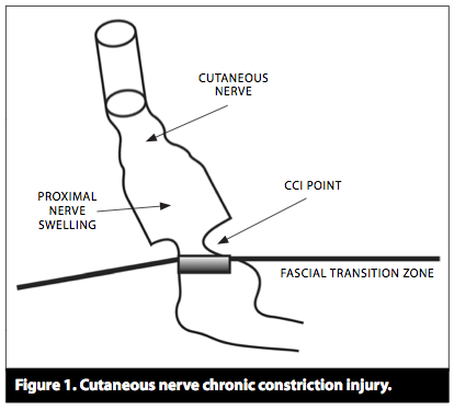
The neuropeptides stored in muscle nociceptors are released not only when peripheral stimuli activate the nerve endings, but also when spinal nerves are compressed. In this type of neuropathic pain, action potentials are generated at the site of compression and spread not only centripetally, i.e., toward the central nervous system, but also centrifugally, i.e., toward the nociceptive endings, where they induce the release of vasoactive neuropeptides. In this way, neurogenic inflammation comes about, characterized by hyperemia, edema, and the release of inflammatory mediators (8). The inflammatory mediators sensitize the muscle nociceptors and thereby increase neuropathic pain.

The sensitization of the muscle nociceptors by endogenous mediators such as BK and PGE2 is one of the reasons why patients with muscle lesions suffer from tenderness to pressure on the muscle, and from pain on movement or exercise. It is also the reason why many types of muscle pain respond well to the administration of non-steroidal anti-inflammatory drugs (NSAID), which block prostaglandin synthesis.

An influx of nervous impulses from muscle nociceptors into the spinal cord increases the excitability of posterior horn neurons to a greater extent than one from cutaneous nociceptors (9).

Two main mechanisms underlie the overexcitability of spinal nociceptive neurons:

A structural change of ion channels, rendering them more permeable to Na+ and Ca2+, is the short-term result of an influx of nociceptive impulses into the spinal cord. Among other effects, this causes originally ineffective («silent» or «dormant») synapses to become effective.

A change of gene transcription in the neuronal nucleus, leading to a modification of synthetic processes, causes new ion channels to be synthesized and incorporated into the nerve cell membrane. The long-term result of central sensitization is a nociceptive cell whose membrane contains a higher density of ion channels that are also more permeable to ions. This explains the hyperexcitability of the cell. Glial cells, too, particularly microglia, can contribute to the sensitization of central neurons by secreting substances such as tumor necrosis factor a (TNF-a) (8).

The increased excitability of spinal neurons and the spread of excitation within the CNS are the first steps in the process of chronification of muscle pain. The endpoint of chronification consists of structural remodeling processes in the CNS that open up new pathways for nociceptive information and cause pain to persist over the long term. Patients with chronic muscle pain are difficult to treat, because the functional and structural changes in the CNS need time to regress. The fact that not all muscle pain becomes chronic implies that chronification requires not only the mechanisms just discussed, but also other ones, e.g., a genetic predisposition.

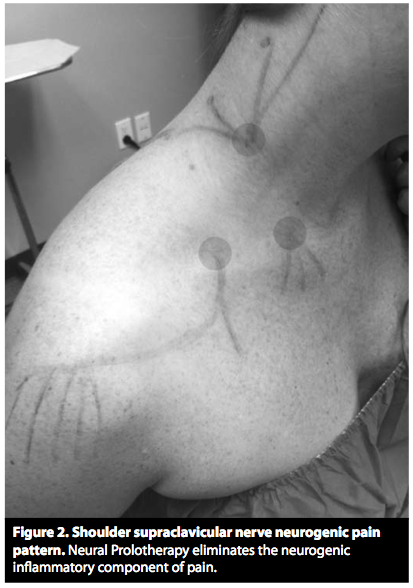
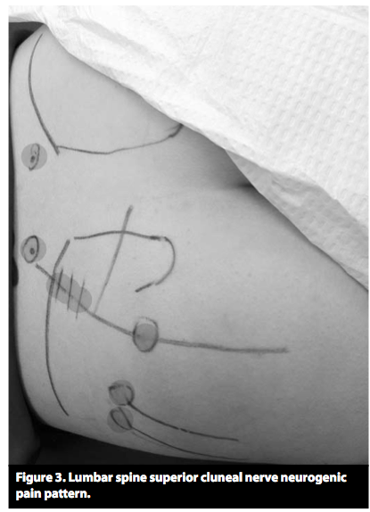
Pain arising in muscle is more likely to be referred pain than pain arising in the skin. Referred pain is pain that is felt not (only) at its site of origin, but at another site some distance away. A possible mechanism of referred pain is the spread, within the spinal cord, of excitation due to the muscle lesion (9) (figures 2 and ​and3).3). As soon as the excitation reaches sensory posterior horn neurons that innervate an area beyond the site of the original muscle lesion, the patient feels referred pain in that area, even though none of the nociceptors in it are activated (13).


An example is shown in figure 3: a stimulus delivered to the myofascial trigger point (MTrP) in the soleus muscle causes only mild local pain, while the patient feels more severe (referred) pain in the sacroiliac joint. No conclusive answers are yet available to the questions of why muscle pain is more likely than cutaneous pain to be referred, why it is usually not referred to both proximal and distal sites, and why pain referral is often discontinuous. There is, however, a well-known discontinuity of spinal topography between the C4 and T2 dermatomes.

The main reason why pain arises in muscle spasm is muscle ischemia, which leads to a drop in pH and the release of pain-producing substances such as bradykinin, ATP, and H+.

The vicious-circle concept of muscle spasm – muscle pain causes spasm, which causes more pain, etc. – should now be considered obsolete. Most studies have shown that muscle pain lowers the excitability of the α-motor neurons innervating the painful muscle (14) (a «pain adaptation» model) (15).

Muscle spasm can be precipitated by, among other things, pain in another muscle. Thus, a spasm-like increase EMG activity in the trapezius muscle has been described in response to painful stimulation of the biceps brachii muscle (16). Another source of muscle spasms is pathological changes in a neighboring joint. These sources of pain must be deliberately sought.

In a widespread hypothesis on the origin of MTrP’s (19), it is supposed that a muscular lesion damages the neuromuscular endplate so that it secretes an excessive amount of acetylcholine. The ensuing depolarization of the muscle cell membrane produces a contraction knot that compresses the neighboring capillaries, causing local ischemia. Ischemia, in turn, leads to the release of substances into the tissue that sensitize nociceptors, accounting for the tenderness of MTrP’s to pressure. Substances of this type have been found to be present within the MTrP’s of these patients (20). This supposed mechanism leaves many questions unanswered but is currently the only comprehensive hypothesis on the origin of MTrP’s.

Patients with MTrP’s often have pain in three locations:

  • at the site of the MTrP itself,
  • at the origin or insertion of the affected muscle, because of pulling by the muscle fibers that have been stretched by the contraction knots,
  • and referred pain outside the MTrP (figure 3).

Because the MTrP is cut off from its blood supply by compression of the local microcirculation, oral NSAID’s are not very effective against TrP pain.

Ukjent sin avatar

Ny behandlingsform mot hodepine og nakkespenninger på Verkstedet

Muskelspenninger helt øverst i nakken bidrar til mange problemer. F.eks. hodepine, spenningsmigrene, kjevespenning, nakkeplager, bevegelsessmerter i nakken, dårlig søvn, m.m.. Spesielt smerter i panna og tinningene har ofte utgangspunkt i området øverst i nakken.

På Verkstedet har vi nå en ny behandlingsform som løser opp dette problemområdet på en svært behagelig og overraskende effektiv måte, og med en eksepsjonelt elegant forklaringsmodell som er på vei til å revolusjonere forståelsen av smertebehandling verden over.

I området som kalles Occiput helt øverst i nakken har vi mange muskler som styrer hodets balanse i alle vinkler. Muskel- og leddterapeuter(fysio, kiro, osteo, massører, osv) tenker vanligvis at det er muskelspenningene som er problemet og at det gjør vondt fordi musklene er stive og inneholder triggerpunkter, eller fordi et ledd er låst. Men med denne nye behandlingsformen innser vi at det er nervesystemet som har problemer, ikke musklene eller leddene. Musklene og leddene gjør bare det nervesystemet befaler. Og det er nervesystemet helt ytterst i huden som reagerer på trykk, IKKE muskelene eller bindevevet. Når vi trykker på en muskel eller et triggerpunkt så er det altså ikke trykket på muskelen du kjenner, men trykket på nervene helt ytterst i huden.

Når vi endelig innser at det er de sensoriske nervene rett under huden som reagerer på trykk og opplevelsen av smerte eller stråling ut panna og tinningene, så kan vi også behandle disse direkte.

I konvensjonell medisin har man skjønt at det er nervetrådene som har problemer. Når det har blitt et seriøst problem kaller de det Trigeminusnevralgi eller Occipetal Nevralgi. Men her behandles disse nervene med f.eks. nedfrysing, bedøvelse, Botox eller avbrenning. I forhold til den nye behandlingformen vi har tatt inn på Verkstedet er dette unødvendig smertefulle og inngripende behandlingsmodeller. Og det værste av alt, de er dyre og har ikke spesielt god effekt heller.

Forskere har også sett at om man bedøver huden når man er støl etter trening, så forsvinner smerten. Selv når man er støl, hvor det kjennes ut som at man har vondt inni muskelen og det er vondt å bevege muskelen, så er det egentlig i nervene helt ytterst i huden som bidrar til smerteopplevelsen. Dette er ikke lett å forstå fordi det er ikke det vi har lært, og det er ikke slik det kjennes ut. Vi har lært at muskelsmerter sitter i musklene, og vi kan verifisere det ved at det «kjennes ut» som at det sitter i musklene. Men som forskerne her har påvist, smerten opprettholdes egentlig i nervetrådene i huden.  https://mariusblomstervik.no/2013/07/14/c-tactile-fibers-contribute-to-cutaneous-allodynia-after-eccentric-exercise/

Med vår nye behandlingsmetode får du en umiddelbar release av muskelspenninger og smerte øverst i nakken. Ikke fordi vi masserer hardt, bedøver eller brenner av nerver, men fordi vi gir huden en behaglig og mild strekk som åpner opp for de minste nervetrådene i huden. De får mer blodsirkulasjon, mer plass, mer næring, og en behagelig stimuli som demper smerte i løpet av noen få minutter.

I forskning regner man at en smertereduksjon på 2 poeng i en 10-poeng skala er «statistisk signifikant». Klienter rappoerterer en umiddelbar reduksjon på 6-8 poeng med denne behandlingsformen. Det er ganske radikalt.

Og det er svært overraskende at noe så enkelt og noe så behagelig kan gi en så stor endring i smerteopplevelsen. Selv de som er vandt til behandling hvor «vondt skal vondt fordrive» lar seg overraske av effekten i dette behandlingskonseptet.

Behandlingsformen kalles DermoNeuroModulation. Legg merke til dette navnet. Dette er begynnelsen på en revolusjon i behandling av smertetilstander. Dermo betyr huden, Neuro betyr nerver, Modulation betyr å endre. Altså, vi endrer nervesystemets respons med behandling av hudens nerver.

Varigheten av forbedringen er avhengig av mange faktorer, bla. ernæring, trening, stressreduksjon og alvorlighetsgraden man kommer med i utgangspunktet. For de fleste gir det en radikal bedring allerede etter første gang, og vanligvis trenger man 2-4 ganger for å få de mest solide resultatene. Noen trenger mer, noen trenger mindre, men ALLE blir overrasket over effekten.

Ta kontakt om du ønsker å se om denne behandlingsformen kan hjelpe for din hodepine eller nakkespenninger. Trykk her for online bestilling 

Ukjent sin avatar

Entrapment Neuropathies in the Upper and Lower Limbs: Anatomy and MRI Features

Nevner viktige steder nerver kommer i klem.

http://www.hindawi.com/journals/rrp/2012/230679/

Although nerves may be injured anywhere along their course, peripheral nerve compression or entrapment occurs more at specific locations, such as sites where a nerve courses through fibroosseous or fibromuscular tunnels or penetrates muscles [2, 3].

Figure 1: The drawing shows anatomy of the suprascapular nerve from the posterior view. Note the nerve courses through the suprascapular notch (open arrow) and spinoglenoid notch (curved arrow). SSN: suprascapular nerve, SS: supraspinatus muscle, IS: infraspinatus muscle.

Suprascapular nerve compression or entrapment, known as suprascapular nerve syndrome, can occur as a result of trauma, an anomalous or thickened transverse scapular ligament, or extrinsic compression by a space-occupying lesion [7, 8], commonly a ganglia cyst or soft tissue tumor. Compression or entrapment at the suprascapular notch leads to supraspinatus and infraspinatus muscle denervation (Figure 2), whereas more distal entrapment at the spinoglenoid notch may present with isolated involvement of the infraspinatus muscle (Figure 3). Patients may present with poorly localized pain and discomfort at the back of the shoulder or the upper back, as well as weakness when raising the arm.


Figure 4: The drawing shows the axillary nerve within the quadrilateral space from a posterior view. AN: axillary nerve, Tm: teres minor muscle, Tr: long head of the triceps, TM: teres major muscle, H: humerus, D: deltoid muscle.

Clinical manifestations include poorly localized shoulder pain and paresthesias in the affected arm in a nondermatomal distribution. The diagnosis can be difficult since clinical symptoms may be confused with rotator cuff pathology or other shoulder joint-related abnormalities [9].


Figure 6: The drawing provides an anterior view of the course of the radial nerve at the elbow. Posterior interosseous nerve (PIN) entrapment may occur due to prominent radial recurrent artery (RRA), medial edge of the extensor carpi radialis brevis (ECRB), and proximal edge of the supinator muscle (SP) (arcade of Frohse). RN: radial nerve, SRN: superficial radial nerve.

The radial nerve is predisposed to injury and entrapment at several locations along its course, which include the radial nerve in the spiral groove of the humerus (spiral groove syndrome) above the elbow joint, where the PIN travels through the radial tunnel, and the superficial branch of the radial nerve where it crosses over the first dorsal wrist compartment (Wartenberg’s syndrome).

Compression or entrapment of the PIN in the radial tunnel may yield two different clinical presentations: posterior interosseous nerve syndrome and radial tunnel syndrome.
In patients with posterior interosseous nerve syndrome, the clinical presentation includes motor deficits of the extensor muscle group without significant sensory loss.
Patients with radial tunnel syndrome, on the other hand, typically present with pain over the proximal lateral forearm [12, 13], which can be caused by acute trauma, masses, and compression from adjacent structures.


Figure 8: The drawing demonstrates the course of the ulnar nerve from posterior view at the elbow. Note the nerve travels deep to the flexor carpi ulnaris muscle (FCU) beneath the arcuate ligament (AL).

Compressive or entrapped ulnar nerve neuropathies include cubital tunnel syndrome and Guyon’s canal syndrome.

Cubital tunnel syndrome is the second most common peripheral neuropathy of the upper extremity. It may be caused by abnormal fascial bands, subluxation, or dislocation of the ulnar nerve over the medial epicondyle, trauma, or direct compression by soft tissue masses. Clinical symptoms include a sensory abnormality of the ulnar hand and weakness of the flexor carpi muscle group of the 4th and 5th fingers.


Figure 12: The drawing of the median nerve shows that it courses along the anterior elbow, through the two heads of the pronator teres muscle (stars), and into the forearm beneath the edge of the fibrous arch of the flexor digitorum sublimis (open arrow).

Median nerve compression or entrapment neuropathies include pronator syndrome, anterior interosseous syndrome, and carpal tunnel syndrome.

Clinical findings include pain and numbness of the volar aspect of the elbow, forearm, and wrist without muscle weakness.


Figure 14: The drawing shows the proximal course the sciatic nerve passing inferior to the piriformis muscle (PS). SG: superior gemellus muscle.

Sciatic nerve entrapment may occur in the hip region and less commonly in the thigh, and clinical presentations are based upon the level of injury [3]. Sciatic neuropathy may result from conditions such as fibrous or muscular entrapment, vascular compression, scarring related to trauma (Figure 15) or radiation, tumors (Figure 16), and hypertrophic neuropathy [3, 17, 18].


Figure 17: Sagittal oblique projection of the knee illustrates the common peroneal nerve (CPN) arising from the sciatic nerve (SN) at the level of popliteal fossa. It travels around the fibular head deep to the origin of the peroneus longus muscle (PL). TN: tibial nerve.

The etiologies of common peroneal neuropathy may include idiopathic mononeuritis, intrinsic and extrinsic space-occupying lesions including an intraneural ganglion cyst (Figure 18) [21], or traumatic injury of the nerve, especially related to proximal fibular fractures [22]. Clinically, patients may experience pain at the site of entrapment with foot drop and a slapping gait [17, 23].


Figure 19: The drawing of the medial aspect of the ankle showing the course of the tibial nerve (TN) and its branches, the medial calcaneal nerve (MCN), and medial and lateral plantar nerves (MPN and LPN), passing through the tarsal tunnel. FR: flexor retinaculum.

Common etiologies include posttraumatic fibrosis due to fracture, tenosynovitis, ganglion cysts (Figure 20), space-occupying lesions, and dilated or tortuous veins. Most patients with tarsal tunnel syndrome have burning pain and paresthesia along the plantar foot and toes.

Ukjent sin avatar

Neural Prolotherapy


Denne artikkelen er om en behandlingsform som sprøyter inn dextrose rett under huden for å stimulere nervetrådene der. Den har mange gode forklaringsmodeller om hva som skjer i nervene rett under huden. Nevner bla anterograd og retrograd nervesignaler i C-fibrene. Og Hiltons Law, som er et svært interessant konsept: nervene som går til et ledd går også til musklene som beveger leddet og huden over muskelens feste. Viser til at dextrose hemmer betennelse i nervene, men dette er et vanskelig konsept ved f.eks. diabetisk nevropati hvor hyperglycemi er noe av årsaken til nerveskaden i utgangspunktet. Dog hyperglycemi påvirker blodsirkulasjonen først og fremst.

http://www.orthohealing.com/wp-content/uploads/2011/10/Neural_prolotherapy.pdf

paThology oF NEUrogENiC iNFlaMMaTioN
The pathology of neurogenic inflammation is well established.1, 2, 16 Ligaments, tendons and joints have TRPV1-sensitive C pain fiber innervation. Dr. Pybus explains that the C pain fibers transmit the “deep pain” often seen with osteoarthritis.14 “When these C pain fibers are irritated anywhere along their length they will transmit ectopic impulses in both forward (prodromic) and reverse (antidromic) direction.”14 The forward direction of the nerve signal will cause pain perception as the signal travels through the posterior root ganglia up to the brain. You will also have a local reflex action from the spinal cord ventral horn cells out to the muscle fibers, which will cause a reflex muscle spasm.14 The reverse (antidromic) signal will travel to the blood vessels where substance P is released causing swelling and pain. The nerves themselves also have a nerve supply called the Nervi Nervorum (NN).2 In a pathological state, the NN can release substance P (Sub P) and Calcitonin Gene Related Peptide (CGRP) onto these C pain fibers.11 Sub P and CGRP are known to cause pain, swelling of the nerve and surrounding tissue.7

Dr. Lyftogt discussed in his recent Neural Prolotherapy meeting that “Cutaneous nerves pass through many fascial layers on their way to the spine. When there is neurogenic swelling at the Fascial Penetration Zone, a Chronic Constriction Injury (CCI) occurs. The CCI points will inhibit flow of Nerve Growth Factor (NGF).8, 7 Proper flow of NGF is essential for nerve health and repair.”3 (See Figure 1.)

Skjermbilde 2013-08-06 kl. 08.59.43

There are two major ways that the fascial penetration point can affect a nerve. Trauma to a nerve will cause edema to travel proximal and distal to the injury. When this swelling reaches the fascial penetration points this can cause a self- strangulation of the nerve and decrease nerve growth factor flow.16, 17 Morton’s neuroma is a clinical example of this.17

Dr. Pybus has also suggested that a change in fascial tension from repetitive muscle dysfunction can also cause a CCI point.15, 17

Another critical concept in NPT is what is called Bystander disease.9, 17 Bystander disease helps explain how superficial nerve pathology can affect deeper anatomic structures.9 This is based on Hilton’s law. Hilton’s law states: the nerve supplying a joint also supplies both the muscles that move the joint and the skin covering the articular insertion of those muscles.9 An example: The musculocutaneous nerve supplies the elbow with pain and proprioception as it is the nerve supply to the biceps brachii and brachialis muscles, as well as the skin close to the insertion of these muscles.17 Hilton’s Law arises as a result of the embryological development of humans.

This concept of Hilton’s law coupled with the idea of anterograde and retrograde axonal flow of neurodegenerative peptides,17 can help explain the wide reaching affects of NPT on pain control.

Glucose responsive nerves have been demonstrated throughout the nervous system.4, 5, 6 One proposed mechanism of action suggests that dextrose binds to pre synaptic calcium channels and inhibits the release of substance P and CGRP, thereby decreasing neurogenic inflammation. This allows normal flow of nerve growth factor and subsequent nerve repair and decreased pain.7

Skjermbilde 2013-08-06 kl. 08.59.48

Skjermbilde 2013-08-06 kl. 09.00.00

1 Geppetti, et al. Neurogenic Inflammation. Boca Raton: Edited CRC Press; 1996. Chapter 5, Summary; p.53-63.

2 Marshall J. Nerve stretching for the relief or cure of pain. The Lancet.1883;2:1029-36.

8 Bennett GJ, et al. A peripheral mononeuropathy in rat that produces disorders of pain sensation like those seen in man. Pain. 1988;33(1):87-107.

9 Hilton J. On rest and Pain. In Jacobesen WHA(ed): On Rest and Pain, 2nd edition, New York: William Wood & company, 1879.

Ukjent sin avatar

Scratch collapse test for evaluation of carpal and cubital tunnel syndrome.

Viser hvilken klinisk relevanse scratch collapse test har for å finne hvor nerver er i klem.

http://www.ncbi.nlm.nih.gov/pubmed/18984333

For the new test, the patient resisted bilateral shoulder external rotation with elbows flexed. The area of suspected nerve compression was lightly «scratched,» and then resisted shoulder external rotation was immediately repeated. Momentary loss of shoulder external rotation resistance on the affected side was considered a positive test.

For carpal tunnel syndrome, sensitivities were 64%, 32%, and 44% for the scratch collapse test, Tinel’s test, and wrist flexion/compression test, respectively. For cubital tunnel syndrome, sensitivities were 69%, 54%, and 46% for the scratch collapse test, Tinel test, and elbow flexion/compression test, respectively. The scratch collapse test had the highest negative predictive value (73%) for carpal tunnel syndrome. Tinel’s test had the highest negative predictive value (98%) for cubital tunnel syndrome.

The scratch collapse test had significantly higher sensitivity than Tinel’s test and the flexion/nerve compression test for carpal tunnel and cubital tunnel syndromes. Accuracy for this test was 82% for carpal tunnel syndrome and 89% for cubital tunnel syndrome.

Mer utfyllende studie om Scratch Collapse her: http://www.ncbi.nlm.nih.gov/pmc/articles/PMC2880669/

Though the exact mechanism of the scratch collapse test is unknown, we believe it may represent a gross physical manifestation of the “cutaneous silent period.” This EMG-demonstrated phenomenon is observed following noxious stimuli. A brief pause of voluntary muscle contraction is demonstrated following stimulation of a cutaneous nerve [24]. The scratch collapse yields a similar reflex response. We propose that as the nervi-nervorum at the site of neuritis are stimulated, an ipsilateral central inhibition is transiently activated. It is not surprising that this response would be most robust at the focus of the neuritis.

The scratch collapse examination shares several features with the cutaneous silent period. Both phenomena occur after a noxious stimulus, are very resistant to habituation, are able to override voluntary muscle contraction, and result in a deferment in resistance in a pattern that corresponds to the withdrawal of the extremity into a position of protection (e.g., in this case, internally rotating the arms in against the body) [911131617]. From an evolutionary standpoint, such a reflex would be important in survival.

The test offers an advantage over these other tests in that it appears to precisely localize the site of nerve compression.

Ukjent sin avatar

Undervisning om nervekompresjon nevropati og kirurgi i armen

Nervekompresjon begynner i en mild variant, hvor myelinlaget rundt nervene fortsatt er tykt og fint. Blodsirkulasjonen hemmes. Smerter og paraestesier kommer og går. Tinels tegn er negativt tidlig i progresjonen. Scratch-Collapse Test viser hvor i nervebanen det er kompresjonproblemer(f.eks. doublecrush syndrome).

Blir moderat, hvor myelinlaget blir tynt. Det blir hevelse i nerven og bindevevet blir tykkere i området. Smerter er konstante og musklene svekkes. Det tar 3-4 mnd å bygge opp myelinlaget igjen når kompresjonen er rettet opp.

Og alvorlig, hvor myelinlaget er borte. Nervetrådene forvinner mer og mer. Atrofi og nummenhet i musklene. Når kompresjonen er borte repareres nerven ca.3 cm i måneden.

http://prezi.com/mjuaxe0cwwbr/?utm_campaign=share&utm_medium=copy&rc=ex0share