Ukjent sin avatar

Breath-holding and its breakpoint

Alt om hva som gjør at vi ikke greier å holde pusten lenge.

http://ep.physoc.org/content/91/1/1.long

This article reviews the basic properties of breath-holding in humans and the possible causes of the breath at breakpoint. The simplest objective measure of breath-holding is its duration, but even this is highly variable. Breath-holding is a voluntary act, but normal subjects appear unable to breath-hold to unconsciousness. A powerful involuntary mechanism normally overrides voluntary breath-holding and causes the breath that defines the breakpoint. The occurrence of the breakpoint breath does not appear to be caused solely by a mechanism involving lung or chest shrinkage, partial pressures of blood gases or the carotid arterial chemoreceptors. This is despite the well-known properties of breath-hold duration being prolonged by large lung inflations, hyperoxia and hypocapnia and being shortened by the converse manoeuvres and by increased metabolic rate.

Breath-holding has, however, two much less well-known but important properties. First, the central respiratory rhythm appears to continue throughout breath-holding. Humans cannot therefore stop their central respiratory rhythm voluntarily. Instead, they merely suppress expression of their central respiratory rhythm and voluntarily ‘hold’ the chest at a chosen volume, possibly assisted by some tonic diaphragm activity. Second, breath-hold duration is prolonged by bilateral paralysis of the phrenic or vagus nerves. Possibly the contribution to the breakpoint from stimulation of diaphragm muscle chemoreceptors is greater than has previously been considered. At present there is no simple explanation for the breakpoint that encompasses all these properties.

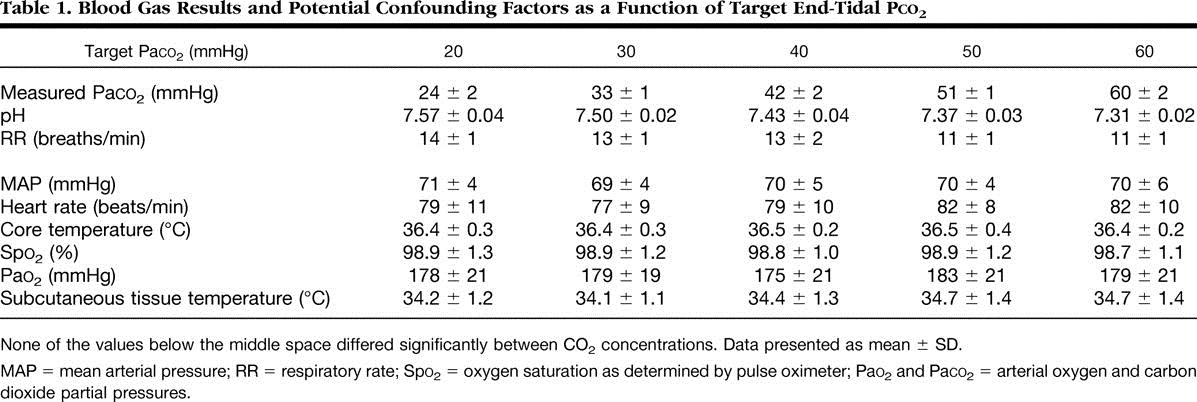
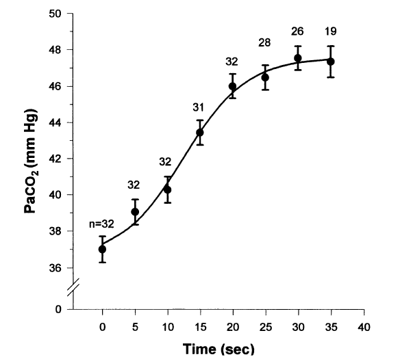
At breakpoint from maximum inflation in air, the Pa/etO2 is typically 62 ± 4 mmHg and the PetCO2 is typically 54 ± 2 mmHg (n = 5; Lin et al. 1974), and the longer the breath-hold the more they change.

Nunn (1987) estimates that consciousness in normal adults is lost at PaO2 levels below ∼27 mmHg and PaCO2 levels between 90 and 120 mmHg.

This is because the rhythmic EMG and negative pressure waves occur simultaneously, because their frequency and amplitude are within the respiratory range and because they increase as CO2 levels rise towards the end of the breath-hold. This CO2 rise would stimulate the central respiratory rhythm.

Ukjent sin avatar

Hypercapnia Improves Tissue Oxygenation

Denne viser hvordan økt CO2 øker oksygenering og blodsirkulasjon i huden og i vevet. Studien er gjort på individer i narkose og med assistert pust med konstant volum på 10ml/kg og pustefrekvens mellom 11 og 14.

http://journals.lww.com/anesthesiology/Fulltext/2002/10000/Hypercapnia_Improves_Tissue_Oxygenation.9.aspx

Background: Wound infections are common, serious, surgical complications. Oxidative killing by neutrophils is the primary defense against surgical pathogens and increasing intraoperative tissue oxygen tension markedly reduces the risk of such infections. Since hypercapnia improves cardiac output and peripheral tissue perfusion, we tested the hypothesis that peripheral tissue oxygenation increases as a function of arterial carbon dioxide tension (Paco2) in anesthetized humans.

Methods: General anesthesia was induced with propofol and maintained with sevoflurane in 30% oxygen in 10 healthy volunteers. Subcutaneous tissue oxygen tension (Psqo2) was recorded from a subcutaneous tonometer. An oximeter probe on the upper arm measured muscle oxygen saturation. Cardiac output was monitored noninvasively. Paco2 was adjusted to 20, 30, 40, 50, or 60 mmHg in random order with each concentration being maintained for 45 min.
Results: Increasing Paco2 linearly increased cardiac index and Psqo2: Psqo2 = 35.42 + 0.77 (Paco2), P < 0.001.
Conclusions: The observed difference in PsqO2 is clinically important because previous work suggests that comparable increases in tissue oxygenation reduced the risk of surgical infection from −8% to 2 to 3%. We conclude that mild intraoperative hypercapnia increased peripheral tissue oxygenation in healthy human subjects, which may improve resistance to surgical wound infections.
co2 og pustefrekvens
co2 og blodsirkulasjon og oksygenering
This hypercapnia-induced increase in cardiac output results in higher tissue oxygen pressure. In the current study Psqo2 went from 58 to 74 mmHg with only a 20-mmHg increase in Paco2. This increase in Psqo2 is likely to be clinically important because it is associated with a substantial reduction in the risk of surgical wound infection. 11 These results suggest that maintaining slight hypercapnia is likely to reduce the risk of surgical wound infection. Carbon dioxide management thus joins the growing list of anesthetic factors that do or are likely to influence the risk of wound infection.

Hypercapnia appears to provide other benefits as well. 35 For example, hypercapnia and hypercapnic acidosis decrease ischemia–reperfusion injury by inhibiting xanthine oxidase in an in vitro model of acute lung injury. 36 Hypercapnia similarly improves functional recovery and coronary blood flow during hypercapnic acidosis in an isolated blood-perfused heart model. 37 Furthermore, small tidal volume ventilation (associated with mild hypercapnia) and permissive hypercapnia have been shown to improve the outcome of patients with acute respiratory distress syndrome as a result of decreased mechanical stretch of the diseased pulmonary tissues. 38,39
Hypercapnia also increases cerebral blood flow and decreases cerebrovascular resistance through dilation of arterioles whereas hypocapnia does the opposite. 40,41 In a recent study, hyper- and hypocapnia were shown to influence brain oxygen tension in swine during hemorrhagic shock 42; hyperventilation and the resulting hypocapnia (15–20 mmHg) decreased cerebral oxygen pressure a further 56%. Hypercapnia has been utilized clinically to improve cerebral perfusion during carotid endarterectomy 43,44 and for emergency treatment of retinal artery occlusion. 45
Ukjent sin avatar

Arterial Blood Gas Changes During Breath-holding From Functional Residual Capacity

Endelig en studie som viser direkte hva som skjer med CO2 når vi holder pusten etter utpust. Her kaller de det etter «functional residual capacity» (wikipedia), altså etter en normal og passiv utpust. Denne studien viser først og fremst hva som skjer i løpet av én enkelt pustehold. De nevner også at det tar 5-10 sekunder etter innpust igjen før CO2 nivet begynner å synke. Så i metablsk pust (RecoveryBreathing) hvor vi puster 10sek inn/ut og har 10 sek pause bør CO2 lett kunne stabiliseres på et høyere nivå enn normalt.

http://www.ncbi.nlm.nih.gov/pubmed/8874252

Hele Studien: http://journal.publications.chestnet.org/data/Journals/CHEST/21737/958.pdf

Breath-holding serves as a model for studying gas exchange during clinical situations in which cessation of ventilation occurs. We chose to examine the arterial blood gas changes that occurred during breath-holding, when breath-holding was initiated from functional residual capacity (FRC) while breathing room air. Eight normal subjects who had a radial artery catheter placed for another study were taught to breath-hold on command from FRC. FRC was determined using respiratory inductance plethysmography. Arterial blood gas specimens were obtained at 5-s intervals until the termination of breath-holding. The average breath-holding time (+/-SD) was 35 (+/-10 s). The PaO2, PaCO2, and pH values were plotted against time and individually fit to logistic equations for each subject. The arterial PaO2 fell by a mean of 50 mm Hg during the first 35 s of breath-holding under these conditions, while the arterial PCO2 rose by a mean of 10.2 mm Hg during the first 35 s and the pH fell by a mean of 0.07 in the first 35 s. The rapid decline in PaO2 is greater than that previously reported using different methods and should be considered in clinical situations in which there is an interruption of oxygenation and ventilation at FRC while breathing room air. The changes in PaCO2 and pH are similar to those previously reported in paralyzed apneic patients.

CO2 etter utpust

We demonstrated during 5 breath-holding runs in which additional arterial specimens were obtained at 5 and 10 s after breath-hold (Fig 6) that the elevated arterial PCO2 did not begin to fall until at least 5 s after breaking from the breath-hold in 1 run and greater than 10 s in 3 other runs. This implies that the removal of the remaining arterial PCO2 by the lungs took longer than 5 s before recirculation from pulmonary capillary blood could lower the arterial PCO2 in the radial artery. The second less significant factor that explains the persistent elevation of arterial PCO2 is the concentrating effect caused by the decreasing LV. The concentrating effect occurs with breath-holding, as more oxygen is removed from the lungs than carbon dioxide is added. As carbon dioxide production continues to occur, the capillary

CO2 fall etter innpust

Ukjent sin avatar

CO2 sin relasjon til pH

Tabell som viser sammenhengen mellom CO2 og pH. CO2 mellom 35-45 er normalt, over eller under dette kaller man det alkalose eller acidose. Med Metabolsk Pust (RecoveryBreathing.com) ønsker vi å få CO2 opp mot 40-50 mmHg. Legg merke til at også HCO3- økes når CO2 økes i blod. Bikarbonat kalles det og er det stoffet som er i Natron.

Klikk for å få tilgang til Oxygenation%20and%20oxygen%20therapy.pdf

TABLE IV

PaCO2 (mm Hg)

pH

HCO3-

15

7.61-7.74

15.3-20.5

20

7.55-7.66

17.7-22.8

30

7.45-7.53

21.0-25.6

40

7.38-7.45

22.8-26.8

50

7.31-7.36

24.1-27.5

60

7.24-7.29

25.1-27.9

70

7.19-7.23

25.7-28.5

80

7.14-7.18

26.2-28.9

90

7.13-7.09

Tabell som viser hvordan O2 og CO2 synker når man kommer opp i høyden.

TABLE V. Gas Pressures at Various Altitudes*

LOCATION

ALT.

PB

FIO2

PIO2

PaCo2

PAO2

PaO2

Sea Level

0

760

.21

150

40

102

95

Cleveland

500

747

.21

147

40

99

92

Denver

5280

640

.21

125

34

84

77

*Pikes’s Peak

14114

450

.21

85

30

62

55

*Mt. Everest

29028

253

.21

43

7.5

35

28

*All pressures in mm Hg; Pike’s Peak and Mt. Everest data from summits

Ukjent sin avatar

Elevated CO2 Levels Cause Mitochondrial Dysfunction and Impair Cell Proliferation

Økt CO2 i cellene gjør at celledeling går tregere. Dette kan fungere også bland kreftceller slik at de deler seg tregere og utvikler seg saktere. De kaller det mitokondrial dysfunksjon i overskriften, men dette er snakk om langvarig hyperkapni opp mot 100 mmHg, langt mer enn hva er mulig å få til med pusteøvelser (50-60 mmHg i korte perioder). Evnen til å kunne øke CO2 i korte perioder vil dermed kunne senke hastigheten på aldringsprosessen

Klikk for å få tilgang til 37067.full.pdf

As shown in Fig. 1, we found a decreased rate of proliferation (assessed as num- ber of cells (Fig. 1, A and B) and BrdU incorporation (Fig. 1, C and D)), which became significant after 3 days of exposure to high levels of CO2. Proliferation was decreased in a dose-depen- dent manner. It is also important to stress that the decreased proliferation was independent of extracellular pH.

Decreased Cell Proliferation during Exposure to High CO2 Levels Is Not Due to Increased Cell Death or Cell Cycle Arrest.

We found that exposure to high CO2 did not result in increased cell death.

We also did not find differences in the distribution of cell cycle phases in cells exposed to high CO2, which rules out a cell cycle arrest as the cause for decreased proliferation. However, the cells had a decreased population doubling rate, indicating that cells exposed to high CO2 have a prolonged cell cycle time. Cells exposed to high CO2 had a slower proliferation rate of 25–30%, pointing to an alteration in cell metabolism, also manifested by decreased oxygen consumption and lower levels of ATP pro- duction (see Fig. 3).

These findings may explain how the cell resists a metabolic stress such as high CO2 by down-regu- lating cell metabolic activity and therefore proliferation and why in diseases such as chronic obstructive pulmonary disease and bronchopulmonary dysplasia there is significant failure to thrive.


Ukjent sin avatar

Insects breathe discontinuously to avoid oxygen toxicity

Veldig spennende artikkel fra Nature om hvordan insekter puster for å unngå oksygenforgftning.

http://www.nature.com/nature/journal/v433/n7025/abs/nature03106.html

http://www.brocku.ca/researchers/glenn_tattersall/research/discussionpapers/Insects%20breathe%20periodically.pdf

The respiratory organs of terrestrial insects consist of tracheal tubes with external spiracular valves that control gas exchange. Despite their relatively high metabolic rate, many insects have highly discontinuous patterns of gas exchange, including long periods when the spiracles are fully closed. Two explanations have previously been put forward to explain this behaviour: first, that this pattern serves to reduce respiratory water loss1, and second, that the pattern may have initially evolved in underground insects as a way of dealing with hypoxic or hypercapnic conditions2. Here we propose a third possible explanation based on the idea that oxygen is necessary for oxidative metabolism but also acts as a toxic chemical that can cause oxidative damage of tissues even at relatively low concentrations. At physiologically normal partial pressures of CO2, the rate of CO2 diffusion out of the insect respiratory system is slower than the rate of O2 entry; this leads to a build-up of intratracheal CO2. The spiracles must therefore be opened at intervals to rid the insect of accumulated CO2, a process that exposes the tissues to dangerously high levels of O2. We suggest that the cyclical pattern of open and closed spiracles observed in resting insects is a necessary consequence of the need to rid the respiratory system of accumulated CO2, followed by the need to reduce oxygen toxicity.

Ukjent sin avatar

Supplementary oxygen for nonhypoxemic patients: O2 much of a good thing?

Nevner alt om hvordan oksygen er skadelig i høye mengder, både i klinisk sammenheng og eller. Bl.a. fordi med høy O2 går CO2 ned og da trekker blodårene seg sammen.

http://www.ncbi.nlm.nih.gov/pmc/articles/PMC3218982/

Abstract

Supplementary oxygen is routinely administered to patients, even those with adequate oxygen saturations, in the belief that it increases oxygen delivery. But oxygen delivery depends not just on arterial oxygen content but also on perfusion. It is not widely recognized that hyperoxia causes vasoconstriction, either directly or through hyperoxia-induced hypocapnia. If perfusion decreases more than arterial oxygen content increases during hyperoxia, then regional oxygen delivery decreases. This mechanism, and not (just) that attributed to reactive oxygen species, is likely to contribute to the worse outcomes in patients given high-concentration oxygen in the treatment of myocardial infarction, in postcardiac arrest, in stroke, in neonatal resuscitation and in the critically ill. The mechanism may also contribute to the increased risk of mortality in acute exacerbations of chronic obstructive pulmonary disease, in which worsening respiratory failure plays a predominant role. To avoid these effects, hyperoxia and hypocapnia should be avoided, with oxygen administered only to patients with evidence of hypoxemia and at a dose that relieves hypoxemia without causing hyperoxia.

… the aim of oxygen therapy should be to increase the delivery of oxygen rather than to reach any arbitrary concentration in the arterial blood.

Hyperoxia marginally increases the arterial blood oxygen content (CaO2), theoretically increasing tissue oxygen delivery (DO2) assuming no reduction in tissue blood flow. However, oxygen causes constriction of the coronary, cerebral, renal and other key vasculatures – and if regional perfusion decreases concomitantly with blood hyperoxygenation, one would have a seemingly paradoxical situation in which the administration of oxygen may place tissues at increased risk of hypoxic stress. Any tissue damage in the course of oxygen administration would plausibly be attributed to the underlying disease process.

Ukjent sin avatar

Respiratory dysregulation in anxiety, functional cardiac, and pain disorders. Assessment, phenomenology, and treatment.

Nevner at hypokapni (lav CO2) er vanlig hos panikkpasienter, de med hjerte/kar problemer og de med smerteproblemer.

http://www.ncbi.nlm.nih.gov/pubmed/11530714

Klikk for å få tilgang til FH%20Wilhelm%20et%20al%20-%20Respiratory%20dysregulation%20review.pdf

Abstract

Respiration is a complex physiological system affecting a variety of physical processes that can act as a critical link between mind and body. This review discusses the evidence for dysregulated breathing playing a role in three clinical syndromes: panic disorder, functional cardiac disorder, and chronic pain. Recent technological advances allowing the ambulatory assessment of endtidal partial pressure of CO2 (PCO2) and respiratory patterns have opened up new avenues for investigation and treatment of these disorders. The latest evidence from laboratories indicates that subtle disturbances of breathing, such as tidal volume instability and sighing, contribute to the chronic hypocapnia often found in panic patients. Hypocapnia is also common in functional cardiac and chronic pain disorders, and studies indicate that it mediates some of their symptomatology. Consistent with the role of respiratory dysregulation in these disorders, initial evidence indicates efficacy of respiration-focused treatment.

Ukjent sin avatar

Primitive, and protective, our cellular oxygenation status?

Om hvordan oksygennivået i mitokondriene er nesten ingen ting, og har vært slik siden tidenes morgen for å beskytte oss mot svingende oksygennivåer i atmosfæren gjennom evlusjonen.

http://www.ncbi.nlm.nih.gov/pubmed/14499489/

The primitive atmosphere where aerobic life started on earth was hypoxic and hypercapnic. Remarkably, an adaptation strategy whereby O2 partial pressure, PO2, in the arterial blood is maintained within a low and narrow range of 1-3 kPa, largely independent of inspired PO2, has also been reported in modern water-breathers. In mammalian tissues, including brain, the most frequently measured PO2 is also in the same low range. Based on the postulate that basic cellular machinery has been established since the early stages of evolution, we propose that this similarity in oxygenation status is the consequence of an early adaptation strategy which, subsequently, throughout the course of evolution, maintained cellular oxygenation in the same low and primitive range independent of environmental changes. Specialized enzymes aimed at protecting cells against O2 toxicity are thought to have appeared very early in evolution but we suggest that preventing high PO2’s is also the simplest and most efficient tool for limiting reactive oxygen species (ROS) production. It could be a cue mechanism to widen our understanding of the ageing process.