Ukjent sin avatar

Different types of slowly conducting afferent units in cat skeletal muscle and tendon.

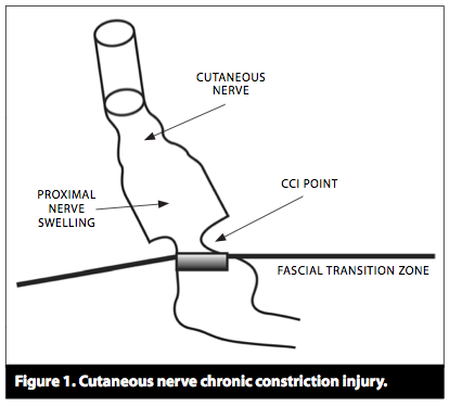
Studie fra 1985 av S.Mense som nevner at det er trykk-sensitive sanseceller i musklene, men at de mest sannsynlig sitter i vevet som omgir muskelen og ikke inni muskelen, siden de ikke reagerer på muskelstrekk eller sammentrekning.

http://m.jp.physoc.org/content/363/1/403.short

Low-threshold pressure-sensitive (l.t.p.) units. Most of these units (ten out of eighteen) responded to touching the receptive field with a soft painter’s brush or a blunt glass rod; they increased their discharge rate upon stimulation with moderate and noxious pressure (Fig. 2). Eight of the l.t.p. units required moderate pressure to be activated;in these cases it was assumed that the mechanosensitive endings were situated more deeply inside the muscle. An observation supporting this assumption was that they did not respond to compression of superficial layers of the muscle with forceps. L.t.p. receptors were relatively insensitive to stretch and contraction; they responded best to forces acting in a direction perpendicular to the long axis of the muscle. Probably, this was due to the fact that many of these receptors were situated in the connective tissue surrounding the tendon and muscle which is not strongly affected by forces building up inside the muscle. (Because of the poor sensitivity of these receptors to the mechanical forces of muscle stretch and contractions,they were not labelled low-threshold mechanosensitive but l.t.p.)

The most frequent type found among group III receptorswas the l.t.p. unit(44%), followed by nociceptive (33%) and contraction-sensitive endings(23%), respectively(Fig.6).

Another feature of l.t.p. units was that they often possessed two receptive fields. In most cases a receptive field with a low mechanical threshold in the distal parts of the muscle or in the tendon was associated with a second receptive field in the proximal or middle third of the muscle, the latter requiring moderate pressure for activation. Out of eighteen l.t.p. units, six(33%) had double receptive fields.

Most of the nociceptive endings had a response behaviour similar to that of cutaneous polymodal nociceptors, in that they responded to both noxious pressure and chemical algesic stimulation.

The physiological significance of the l.t.p. units is not clear. Judging from their response behaviour they would be suited to signal innocuous deformation of muscle and tendon.

In the older literature, there are some reports suggesting the existence of a deep pressure sense in man (Head,Rivers& Sherren, 1905; Lewis & Pochin, 1938; Kellgren & McGowan, 1948), but whether the deep pressure sensations are due to activation of slowly conducting afferent fibres is unknown at present.

Ukjent sin avatar

En del studier relatert til DermoNeuroModulation

En del studier relatert til hudstekk behandlingen, DermoNeuroModulation:

Cutaneous afferents provide information about knee joint movements in humans

 http://jp.physoc.org/content/531/1/289.full

To investigate if proprioceptive information is also provided by skin mechanoreceptor afferents from skin areas related to large joints of postural importance, microneurography recordings were obtained in humans from skin afferents in the lateral cutaneous femoral nerve to study their responses to knee joint movements.

All afferents from fast and slowly adapting low-threshold mechanoreceptors, as well as C mechanoreceptors, responded to manually applied skin stretch. In contrast, the same stimulus elicited, at most, feeble responses in hair follicle receptors.

Qualitative and quantitative analyses of the responses of a subset of afferents revealed that in particular slowly adapting afferents effectively encode both static and dynamic aspects of passively imposed knee joint movements.

A previously undefined type of slowly adapting receptor (SA III) seemed particularly suited for this task whereas this does not seem to be the case for either hair follicle receptors or C mechanoreceptors.

Cortical processing of lateral skin stretch stimulation in humans.

 http://www.ncbi.nlm.nih.gov/pubmed/18574581

Direction discrimination of a moving tactile stimulus requires intact dorsal columns and provides a sensitive clinical test of somatosensory dysfunction

Second somatosensory cortex (S2) was activated in the task as well as no task experiment, and there was no significant difference in cortical activation between the two experiments. Within S2 nearly all subjects had prominent activations in the caudal and superficial part, i.e., in the opercular parietal (OP) area 1.

Tactile directional sensitivity and postural control.

 http://www.ncbi.nlm.nih.gov/pubmed/16143860

Tactile directional sensitivity depends on two different kinds of somatosensory information, i.e. spatiotemporal information and information about friction-induced changes in skin stretch. The objective of this study was to compare the relative contribution to postural control of these two types of information for both glabrous and hairy skin. Postural sway amplitudes and sway paths were recorded, with or without access to tactile and/or visual stabilizing stimuli. Subjects were standing on two types of surface, either solid metal or 50 mm foam plastic.

The results invite speculation that patients with poor directional sensitivity might have reduced postural stability compared with healthy individuals.

Does sympathetic nerve discharge affect the firing of myelinated cutaneous afferents in humans?

http://www.ncbi.nlm.nih.gov/pubmed/15182741

The close relation to blood flow for all types of afferents, and the different responses among SAII afferents, suggest that sympathetically mediated changes in afferent firing properties are indirect, i.e. secondary to changes in the mechanoreceptors’ tissue environment rather than to a direct sympathetic effect on the endings.

Clustering of slowly adapting type II mechanoreceptors in human peripheral nerve and skin.

http://www.ncbi.nlm.nih.gov/pubmed/9549505

At many sites two, sometimes even three, neighbouring SAII units were recorded from the explored nerve fascicle and they had adjacent or even overlapping cutaneous receptive fields.

The neighbouring SAII units were optimally activated by stretching the skin in different directions. Stretching the same skin area in different directions produced different unit recruitment.

Clustered SAII units were often found in sites where Pacinian afferents and skin sympathetic activity were also recorded.

The data do not support the notion that myelinated fibres are randomly organized in peripheral nerve fascicles. Instead, the findings suggest that SAII units tend to be clustered in human peripheral nerves. Furthermore, the response of groups of SAII units to skin stretch suggests that they play a role in proprioception.

Ukjent sin avatar

Functional Anatomy of Muscle – Muscle, Nociceptors and Afferent Fibers

Svært mye interessant om nervesystemets rolle i muskler og smerte.

Spesielt at det er ingen frie nerveender i muskelcellene, men bare i blodkarene i musklene. Derfor reagerer vi med smerte på betennelser og lav pH i blodet, mens trykksensitiviteten kun sitter i huden.

Den nevner at pH sensibiliteten er den viktigste smertebidraget fordi pH synker i de fleste patologiske tilstander, f.eks. hard trening eller skade.

Den nevner at det er mer SP (Substans P, som er relatert til smerte) i huden enn i muskler.

Nevner at frie nerveender ikke går til kapillærer eller muskelceller, bare til arterioler og venuler.

Nevner også innervering av bindevev, og at dette feltet foreløpig er lite studert og oversett. Spesielt viser de til at Toracolumbar Facia (i korsryggen) har størst innervasjon av C-fiber nociceptorer(som inneholder SP) under huden, og litt i multifidene.

En nociceptor er ikke bare en passiv mottaker av impulser, men er også en aktiv deltaker i vevets tilstand når det gjelder betennelser og blodsirkulasjon for de sender nevropetider ut fra doresalhornet til vevet (antidromiske impulser). Altså motsatt vei av reseptor-signalretningen.

CGRP virker vasodilerende, mens SP gjør at blodkarveggenes permeabilitet øker. Når permeabiliteten øker siver det ut proteiner og stoffer som egentlig ikke skal være i vevet, og da økes betennelser og immunsystemets aktivitet. Så det er SP vi ønsker å dempe først og fremst.

http://www.google.com/url?sa=t&rct=j&q=&esrc=s&source=web&cd=1&cad=rja&ved=0CCsQFjAA&url=http%3A%2F%2Fwww.springer.com%2Fcda%2Fcontent%2Fdocument%2Fcda_downloaddocument%2F9783540850205-c1.pdf%3FSGWID%3D0-0-45-935848-p173845615&ei=_GJEUqyUOevn4QSBl4HQDg&usg=AFQjCNEpyWrolHywvUNyw_Cwa8yWTiEUnw&sig2=OJLZ3XrnTalCUUqE-uhCeQ&bvm=bv.53217764,d.bGE

The predominant location of free nerve endings supplied by group IV fibers was the adventitia of arterioles and venules. Surprisingly, muscle fibers themselves did not receive direct innervation by free nerve endings. Group III afferents generated not only free nerve endings but also paciniform corpuscles, whereas group IV fibers terminated exclusively in free nerve endings.The high sensitivity of the free nerve endings to chemical stimuli, particularly to those accompanying inflammatory lesions or disturbances of the microcirculation, may be related to their location on or in the walls of the blood vessels. The finding that the muscle fibers proper are not supplied by free nerve endings (Reinert et al. 1998) may relate to the clinical experience that muscle cell death is usually not painful, at least not if it occurs slowly, as for instance during muscular dystrophy, polymyositis, or dermatomyositis. A different situation is tearing of a muscle fiber bundle, which can be extremely painful. In this condi- tion, many muscle cells are destroyed simultaneously and release their contents (e.g., K+ ions and ATP) in the interstitial space, from where they can diffuse to the next nociceptive endings.
In skeletal muscle, the free nerve endings appear to be distributed quite evenly in the proximodistal direction. At least, in a quantitative evaluation of the inner- vation density by neuropeptide-(SP- and CGRP-) containing fibers, no difference between the proximal and distal portions of the rat gastrocnemius–soleus (GS) muscle was found (Reinert et al. 1998). Therefore, a higher innervation density at the transition zone between muscle and tendon is not a probable explanation for the frequent pain in this region.

However, in the same study the nerve fiber density in the peritendineum (the connective tissue around a tendon) of the rat calcaneal tendon was found to be several times higher than that in the GS muscle. In contrast, the collagen fiber bundles of the tendon tissue proper were almost free of free nerve endings. The high fiber density in the peritendineum may explain the high prevalence of tenderness or pain in the tissue around the tendon and the insertion site. The scarcity of nerve endings in the center of the tendon may relate to the clinical observation that (incomplete) ruptures of the tendon may occur without pain.

Judging from their respon- siveness to pain-producing agents, the following receptor molecules are likely to be relevant for muscle pain and tenderness (Mense and Meyer 1985; Caterina and David 1999; McCleskey and Gold 1999; Mense 2007):

  • Bradykinin (BKN) receptors (B1 and B2). BKN is cleaved from blood plasma proteins when a blood vessel breaks or increases its permeability so that plasma proteins enter the interstitial space. In intact tissue, BKN excites nerve endings by the activation of the BKN receptor molecule B2, whereas under pathological conditions (e.g., inflammation) the receptor B1 is the predominant one (Perkins and Kelly 1993; for a review of receptor molecules mediating the effects of classic inflammatory (pain-producing or algesic) substances, see Kumazawa 1996).
  • Serotonin receptors (particularly 5-HT3). Serotonin (5-hydroxytryptamin, 5-HT) is released from blood platelets during blood clotting. The stimulating effects of serotonin on nociceptive terminals in the body periphery are predomi- nantly mediated by the 5-HT3 receptor (at present, more than 15 different 5-HT receptors are known in the CNS). The serotonin concentrations released in the tissue are usually not sufficient to excite nociceptors directly, but they can sen- sitize them, i.e., make them more sensitive to other pain-producing agents such as BKN.
  • Prostaglandins, particularly prostaglandin E2 (PGE2). Prostaglandins (PGs) are released in a pathologically altered muscle by the enzymatic action of cycloox- igenases. PGE2 binds to a G protein-coupled prostanoid receptor (EP2) in the membrane of the nociceptive ending. Similarly to serotonin, PGE2 sensitizes nociceptors rather than exciting them under (patho)physiological circumstances (Mense 1981).
  • Acid-sensing ion channels (ASICs). ASICs constitute a family of receptor molecules that are sensitive to a drop in pH and open at various pH values. The channel proteins react already to small pH changes, for instance from pH 7.4 to 7.1. This receptor family (for instance ASIC1 and ASIC3) is particularly impor- tant for muscle pain, because almost all pathologic changes in muscle are accom- panied by a drop in tissue pH, e.g., exhausting exercise, ischemia, and inflammation (Immke and McCleskey 2003). In these conditions, the pH of the muscle tissue can drop to 5–6. The proton-sensitive nociceptors may also be of importance for the induction of chronic muscle pain. Repeated intramuscular injections of acidic solutions have been reported to induce a long-lasting hyper- algesia (Sluka et al. 2001).
  • P2X3 receptors. This receptor is a subtype of the purinergic receptors that are activated by ATP and its derivatives (Burnstock 2007; Ding et al. 2000). ATP is the energy-carrying molecule in all cells of the body; accordingly, it is present in every tissue cell. It is released from all tissues during trauma and other pathologic changes that are associated with cell death. For this reason, ATP has been considered a general signal substance for tissue trauma and pain (Cook and McCleskey 2002). ATP is particularly important for muscle pain, because it is present in muscle cells in high concentration (Stewart et al. 1994). When injected into human muscle, ATP causes pain (Mo ̈rk et al. 2003).
  • Transient receptor potential receptor subtype 1 (TRPV1) formerly called VR1. This receptor is one of the most important molecules for the induction of pain. The natural stimulant for the TRPV1 receptor is Capsaicin, the active ingredient of chilli peppers (Caterina and Julius 2001). The receptor is also sensitive to an increase in H+-concentration and to heat, with a threshold of approximately 39C. Its endoge- nous ligands are H+-ions.
  • Other TRP receptors. TRPV4 is a mechanosensitive ion channel that is sensitive to both weak and strong (noxious) intensities of local pressure (Liedtke 2005). It may be the receptor for mediating pain evoked by pinching and squeezing.
  • Tyrosine kinase A (TrkA) receptor. The ligand of this receptor is NGF (Caterina and David 1999). NGF is well-known for its sensitizing action on nociceptors in the body periphery and neurons in the CNS; it is synthesized in muscle, and its synthesis is increased during pathophysiological changes of the muscle (e.g., inflammation, Menetrey et al. 2000; Pezet and McMahon 2006).
  • Glutamate receptors. There is evidence indicating that the NMDA receptor (one of the receptors for glutamate) is present on nociceptive endings in masticatory muscles. Injections of glutamate into the masseter muscle in human subjects induced a reduction in pressure pain threshold which was attenuated by coinjection with ketamine, an NMDA receptor antagonist (Cairns et al. 2006).
Substances exciting muscle nociceptors independent of membrane receptors.
  • Hypertonic saline: injections of NaCl solutions (4.5–6.0%) have long been and still are used to elicit pain from deep somatic tissues (Kellgren 1938; for review, see Graven-Nielsen 2006).
  • Potassium ions: The most likely explanation for the excitatory action of high concentrations of extracellular potassium ions is a depolarization of the membrane potential due to a reduction of the inside–outside potassium gradient (usually the potassium concentration inside the axon is much higher).

DRG cells projecting in a cutaneous nerve have been reported to contain SP, CGRP, and somatostatin (SOM).

In comparison to skin nerves, muscle nerves appear to contain less SP. This finding makes sense, because the vasodilatation and plasma extrava- sation caused by the release of SP and CGRP from free nerve endings (see below) would be dangerous for skeletal muscles, since many of them are surrounded by a tight fascia. Therefore, an SP-induced muscle edema would result in a high increase in interstitial pressure, and could cause muscle necrosis.

In a study on functionally identified DRG cells employing a combination of electrophysiological and immunohistochemical techniques, Lawson et al.(1997) reported that cells terminating in cutaneous nociceptive endings showed a strong tendency to express SP, particularly if they had a slow conduction velocity or a small soma in the DRG. 

The peptides are synthesized in the somas of the DRG or in ganglion cells of cranial nerves. They are transported to both the central and the peripheral terminal of the primary afferent unit.

In a quantitative evaluation of neuropeptide-containing free nerve endings and preterminal axons (both characterized by varicosities) in the GS muscle of the rat, most endings were found around small blood vessels (arterioles or venules), whereas capillaries and the muscle cells proper were not contacted by these end- ings.

Efferent or motor fibers conduct action potentials from the CNS to the periphery; their soma is located in the spinal cord or brainstem and the fibers leave the CNS via the ventral root or cranial nerve motor roots. An exception are postganglionic sympathetic fibers whose cell bodies are mostly located in the sympathetic trunk (e.g., vasomotor fibers that constrict blood vessels).

The nerve to a locomotor muscle in the cat (the lateral GS) is composed of approximately one-third myelinated (720) and two-thirds unmyelinated (2,480) fibers (Table 2.2; Mitchell and Schmidt 1983; Stacey 1969). Nearly one quarter of the myelinated (group III) fibers had nociceptive properties in neurophysio- logical experiments (Mense and Meyer 1985). Of the unmyelinated fibers, 50% are sensory (group IV), and of these, approximately 55% have been found to be nociceptive in the rat (Hoheisel et al. 2005).

Data obtained from one muscle nerve cannot be transferred directly to other muscle nerves, because considerable differences exist between different muscles. For instance, neck muscle nerves of the cat contain unusually high numbers of sensory group III receptors (Abrahams et al. 1984). One possible explanation for these differences is that the muscles have different functions and environmental conditions: in contrast to the neck muscles, which must register the orientation of the head in relation to the body in fine detail, the locomotor hindlimb muscles often have to contract with maximal strength and under ischemic conditions.

In addition to nociceptors, there are other muscle receptors whose function is essential for the understanding of muscle pain:

  • Muscle spindles are complex receptive structures that consist of several specialized muscle fibers (the so-called intrafusal muscle fibers; the name is derived from their location inside the spindle-shaped connective tissue sheath. Accordingly, all the “normal” muscle fibers outside the spindle are “extrafusal” fibers). Muscle spindles measure the length and the rate of length changes of the muscle, i.e., their discharge rate increases with increasing muscle length and with increasing velocity of the length change.
  • Golgi (tendon) organs measure the tension of the muscle. They are arranged in series with the extrafusal muscle fibers; their location is the transition zone between muscle and tendon. The supplying fiber is the Ib afferent, whose structure is identical to the Ia fiber (thick myelin sheath and high conduction velocity). The receptor has a much simpler structure than the muscle spindle; it consists of receptive endings that are interwoven between the collagen fiber bundles of the tendon.
  • Muscle spindles and Golgi organs are proprioceptors, i.e., they measure the internal state of the body.
  • Pacinian corpuscles (PC) and paciniform corpuscles. These receptors do not respond to static pressure; they require dynamically changing mechanical stimuli, and are best excited by vibrations of relatively high frequency (close to 300 Hz; Kandel et al. 2000). The receptive ending is formed like a rod, and covered by several concentric membranes which give the receptor an onion-like appearance in cross-sections.

At present, little information is available about the innervation of fascia. This is an important gap in our knowledge, because fascia is an important component of the musculoskeletal system and likely to contribute to many forms of pain that are subsumed under the label “muscle” pain. One example is low back pain: The thoracolumbar fascia (TF) plays an essential role in body posture and trunk move- ments (Bogduk and Macintosh 1984). It is not only a passive transmitter of mechanical forces of the low back and abdominal muscles but also contractile by itself (Schleip et al. 2005).

In the connective tissue around the superficial lamina of the TF we found many CGRP- and SP-containing free nerve endings. The majority of the fibers were located in the subcutaneous layer, as well as between the fascia and the surface of the multifidus muscle (Fig. 2.8). The SP-positive endings are of particular interest, because they are thought to be nociceptors.
The loose connective tissue around the TF is probably deformed during any trunk movement, and therefore the free nerve endings are strategically situated to sense any disorders in these movements. It is conceivable that overload of the fascia puts mechanical stress and irritation on the endings, and thus may contribute to low back pain.
SP then releases histamine from mast cells, and together with CGRP these agents cause vasodilatation and an increase in vascular permeability of the blood vessels around the active ending. The result is a shift of blood plasma from the intravascular to the interstitial space. Outside the blood vessel, BKN is cleaved from the plasma protein kallidin, serotonin (5-HT) is set free from platelets, and PGs (particularly PGE2) from endothelial and other tissue cells. All these substances sensitize nociceptors. Thus, the main tissue alteration induced by a nondestructive noxious mechanical stimulus is a localized region of vasodilatation, edema, and sensitized nociceptors.
A nociceptor is not a passive sensor of tissue-threatening stimuli; it actively influences the microcirculation and chemical composition of the intersti- tial space around it.
If a noxious stimulus activates only one part of the ending, the action potentials originating in that region of the ending can invade antidromically (against the normal direction of propagation) those branches of the ending that were not excited by the stimulus. These antidromic action potentials release neuropeptides from the unstimulated branches. The whole process is called the axon reflex.  It is assumed to be the reason for the visible wheal and flare around a cutaneous lesion.

The vascular permeability is increased mainly by SP (and by the neurokinins A and B; Gamse and Saria 1985), whereas CGRP is assumed to act primarily as a vasodilator. There is evidence showing that CGRP enhances the plasma extravasation induced by SP and neurokinins A and B, but reduces the vasodilatory action of SP by desensitizing muscle arterioles to the peptide (O ̈ hle ́n et al. 1988).

The area of wheal and flare after a localized damage to the skin – for instance around a needle prick – could be an indicator of the extent of the excited nocicep- tive ending.

The size of the receptive fields (RFs) of cutaneous polymodal nociceptors was found to be less than 2 mm in cat (Bessou and Perl 1969) and 6–32 mm in rabbit (Kenins 1988). A receptive field is that region of the body from which a receptive ending (or a central sensory neuron) can be excited. The above figures are larger than the reported length of the branches of a nociceptor ending (a few hundred mm; Stacey 1969).

The release of SP, CGRP, neurokinin A, and other agents from nociceptors is the central factor in the cascade of events that lead to neurogenic inflammation in the periphery (Lembeck and Holzer 1979). Neurogenic inflammation is characterized by tissue edema and infiltration by immune cells, i.e., it exhibits the major histo- logical signs of a (sterile) inflammation. It develops whenever action potentials are generated not at the receptive ending but somewhere along the course of primary afferent units (spinal nerve or dorsal root). The action potentials propagate both to the CNS (causing pain) and to the peripheral ending (causing release of neuropep- tides and neurogenic inflammation). The published data indicate that vasodilatation can be elicited by antidromic stimulation of both Ad- and C fibers, but increase in vascular permeability and plasma extravasation by stimulation of C fibers only.

Neuropathies and radiculopathies and other pathological conditions that are asso- ciated with antidromic activity in sensory nerve fibers are examples of such events (Marchand et al. 2005). Neurogenic inflammation is likely to increase the dysesthe- sia and pain of patients suffering from neuropathies.

Inflammatory disorders are usually accompanied by sensitization of peri- pheral nociceptors, which is one source of inflammatory pain (for details, see Chap. 3). 

Ukjent sin avatar

Ny behandlingsform mot hodepine og nakkespenninger på Verkstedet

Muskelspenninger helt øverst i nakken bidrar til mange problemer. F.eks. hodepine, spenningsmigrene, kjevespenning, nakkeplager, bevegelsessmerter i nakken, dårlig søvn, m.m.. Spesielt smerter i panna og tinningene har ofte utgangspunkt i området øverst i nakken.

På Verkstedet har vi nå en ny behandlingsform som løser opp dette problemområdet på en svært behagelig og overraskende effektiv måte, og med en eksepsjonelt elegant forklaringsmodell som er på vei til å revolusjonere forståelsen av smertebehandling verden over.

I området som kalles Occiput helt øverst i nakken har vi mange muskler som styrer hodets balanse i alle vinkler. Muskel- og leddterapeuter(fysio, kiro, osteo, massører, osv) tenker vanligvis at det er muskelspenningene som er problemet og at det gjør vondt fordi musklene er stive og inneholder triggerpunkter, eller fordi et ledd er låst. Men med denne nye behandlingsformen innser vi at det er nervesystemet som har problemer, ikke musklene eller leddene. Musklene og leddene gjør bare det nervesystemet befaler. Og det er nervesystemet helt ytterst i huden som reagerer på trykk, IKKE muskelene eller bindevevet. Når vi trykker på en muskel eller et triggerpunkt så er det altså ikke trykket på muskelen du kjenner, men trykket på nervene helt ytterst i huden.

Når vi endelig innser at det er de sensoriske nervene rett under huden som reagerer på trykk og opplevelsen av smerte eller stråling ut panna og tinningene, så kan vi også behandle disse direkte.

I konvensjonell medisin har man skjønt at det er nervetrådene som har problemer. Når det har blitt et seriøst problem kaller de det Trigeminusnevralgi eller Occipetal Nevralgi. Men her behandles disse nervene med f.eks. nedfrysing, bedøvelse, Botox eller avbrenning. I forhold til den nye behandlingformen vi har tatt inn på Verkstedet er dette unødvendig smertefulle og inngripende behandlingsmodeller. Og det værste av alt, de er dyre og har ikke spesielt god effekt heller.

Forskere har også sett at om man bedøver huden når man er støl etter trening, så forsvinner smerten. Selv når man er støl, hvor det kjennes ut som at man har vondt inni muskelen og det er vondt å bevege muskelen, så er det egentlig i nervene helt ytterst i huden som bidrar til smerteopplevelsen. Dette er ikke lett å forstå fordi det er ikke det vi har lært, og det er ikke slik det kjennes ut. Vi har lært at muskelsmerter sitter i musklene, og vi kan verifisere det ved at det «kjennes ut» som at det sitter i musklene. Men som forskerne her har påvist, smerten opprettholdes egentlig i nervetrådene i huden.  https://mariusblomstervik.no/2013/07/14/c-tactile-fibers-contribute-to-cutaneous-allodynia-after-eccentric-exercise/

Med vår nye behandlingsmetode får du en umiddelbar release av muskelspenninger og smerte øverst i nakken. Ikke fordi vi masserer hardt, bedøver eller brenner av nerver, men fordi vi gir huden en behaglig og mild strekk som åpner opp for de minste nervetrådene i huden. De får mer blodsirkulasjon, mer plass, mer næring, og en behagelig stimuli som demper smerte i løpet av noen få minutter.

I forskning regner man at en smertereduksjon på 2 poeng i en 10-poeng skala er «statistisk signifikant». Klienter rappoerterer en umiddelbar reduksjon på 6-8 poeng med denne behandlingsformen. Det er ganske radikalt.

Og det er svært overraskende at noe så enkelt og noe så behagelig kan gi en så stor endring i smerteopplevelsen. Selv de som er vandt til behandling hvor «vondt skal vondt fordrive» lar seg overraske av effekten i dette behandlingskonseptet.

Behandlingsformen kalles DermoNeuroModulation. Legg merke til dette navnet. Dette er begynnelsen på en revolusjon i behandling av smertetilstander. Dermo betyr huden, Neuro betyr nerver, Modulation betyr å endre. Altså, vi endrer nervesystemets respons med behandling av hudens nerver.

Varigheten av forbedringen er avhengig av mange faktorer, bla. ernæring, trening, stressreduksjon og alvorlighetsgraden man kommer med i utgangspunktet. For de fleste gir det en radikal bedring allerede etter første gang, og vanligvis trenger man 2-4 ganger for å få de mest solide resultatene. Noen trenger mer, noen trenger mindre, men ALLE blir overrasket over effekten.

Ta kontakt om du ønsker å se om denne behandlingsformen kan hjelpe for din hodepine eller nakkespenninger. Trykk her for online bestilling 

Ukjent sin avatar

Sensory innervation of the thoracolumbar fascia in rats and humans.

Studie som viser innervasjon av korsryggbindevev og påpeker at det er kun det ytre laget av bindevevet, det som er helt inn mot huden, som er tettpakket med sensoriske nerver og nociceptive fibre (som utskiller substans P og CGRP, og gir betennelser). De dypere lagene i midten av bindevevet eller ned mot musklene har nesten ingen nerveender eller sansesmuligheter.

http://www.ncbi.nlm.nih.gov/pubmed/21839150

Hele studien i min dropbox.

The subcutaneous tissue and the outer layer showed a particularly dense innervation with sensory fibers. SP-positive free nerve endings-which are assumed to be nociceptive-were exclusively found in these layers. Because of its dense sensory innervation, including presumably nociceptive fibers, the TLF may play an important role in low back pain.


Fig. 1. Structure of the rat thoracolumbar fascia (TLF) close to the spinous processes L4/L5. (a) Transversal section showing the three layers of the TLF (hematoxylin and eosin staining): OL, outer layer with transversely oriented collagen fibers; ML, middle layer composed of collagen fiber bundles oriented diagonally to the long axis of the body; IL, inner layer of loose connective tissue covering the multifidus muscle (muscle). SCT, subcutaneous tissue. (b) PGP 9.5-ir nerve fibers in the layers of the TLF. Black arrows, fibers on passage; open arrows, nerve endings. (c) Mean fiber length of PGP 9.5-ir fibers in the TLF. The great majority of all fibers were located in the outer layer (OL) of the fascia and in the subcutaneous tissue (SCT). White part of the bar: subcutaneous tissue plus outer layer of the TLF; black: middle layer; hatched: inner layer. n, number of sections evaluated.


Fig. 4. Distribution of CGRP and Substance P (SP)-immunoreactive nerve fibers in the TLF. (a) Mean fiber length of CGRP-ir nerve fibers. (b) Mean fiber length of SP-ir nerve fibers. Almost all fibers were found in the outer layer of the fascia and the subcutaneous tissue. The middle layer was free of SP-positive fibers. Gray part of the bars: subcutaneous tissue; white: outer layer of the TLF; black: middle layer; hatched: inner layer. n=number of sections evaluated. (c, d) Distribution of CGRP- (c) and SP-containing receptive free nerve endings (d) expressed as percent of the total number of CGRP- or SP-containing fibers in each layer. For classification as receptive endings, the structures had to exhibit at least three varicosities. SP-containing free nerve endings were restricted to the outer layer of the thoracolumbar fascia and the subcutaneous connective tissue while CGRP-containing free nerve endings were also found in the inner layer of the thoracolumbar fascia.

Og et bilde av de forskjellige bindevevslagene som er nevnt i denne studien.

Our study demonstrates that the rat TLF and the SCT overlying the fascia are densely innervated tissues, and therefore both the TLF and SCT, may play a role in low back pain. Most nerve fibers are located in the OL of the TLF and in the SCT, whereas in the ML nerve fibers are rare. Actually, no SP-ir fibers were found in this layer. Teleologically, the lack of fibers in the ML, particularly those containing SP, makes sense because each move- ment of the body causes shearing forces between the collagen fiber bundles, which might excite nociceptors.

Ukjent sin avatar

Neural Prolotherapy


Denne artikkelen er om en behandlingsform som sprøyter inn dextrose rett under huden for å stimulere nervetrådene der. Den har mange gode forklaringsmodeller om hva som skjer i nervene rett under huden. Nevner bla anterograd og retrograd nervesignaler i C-fibrene. Og Hiltons Law, som er et svært interessant konsept: nervene som går til et ledd går også til musklene som beveger leddet og huden over muskelens feste. Viser til at dextrose hemmer betennelse i nervene, men dette er et vanskelig konsept ved f.eks. diabetisk nevropati hvor hyperglycemi er noe av årsaken til nerveskaden i utgangspunktet. Dog hyperglycemi påvirker blodsirkulasjonen først og fremst.

http://www.orthohealing.com/wp-content/uploads/2011/10/Neural_prolotherapy.pdf

paThology oF NEUrogENiC iNFlaMMaTioN
The pathology of neurogenic inflammation is well established.1, 2, 16 Ligaments, tendons and joints have TRPV1-sensitive C pain fiber innervation. Dr. Pybus explains that the C pain fibers transmit the “deep pain” often seen with osteoarthritis.14 “When these C pain fibers are irritated anywhere along their length they will transmit ectopic impulses in both forward (prodromic) and reverse (antidromic) direction.”14 The forward direction of the nerve signal will cause pain perception as the signal travels through the posterior root ganglia up to the brain. You will also have a local reflex action from the spinal cord ventral horn cells out to the muscle fibers, which will cause a reflex muscle spasm.14 The reverse (antidromic) signal will travel to the blood vessels where substance P is released causing swelling and pain. The nerves themselves also have a nerve supply called the Nervi Nervorum (NN).2 In a pathological state, the NN can release substance P (Sub P) and Calcitonin Gene Related Peptide (CGRP) onto these C pain fibers.11 Sub P and CGRP are known to cause pain, swelling of the nerve and surrounding tissue.7

Dr. Lyftogt discussed in his recent Neural Prolotherapy meeting that “Cutaneous nerves pass through many fascial layers on their way to the spine. When there is neurogenic swelling at the Fascial Penetration Zone, a Chronic Constriction Injury (CCI) occurs. The CCI points will inhibit flow of Nerve Growth Factor (NGF).8, 7 Proper flow of NGF is essential for nerve health and repair.”3 (See Figure 1.)

Skjermbilde 2013-08-06 kl. 08.59.43

There are two major ways that the fascial penetration point can affect a nerve. Trauma to a nerve will cause edema to travel proximal and distal to the injury. When this swelling reaches the fascial penetration points this can cause a self- strangulation of the nerve and decrease nerve growth factor flow.16, 17 Morton’s neuroma is a clinical example of this.17

Dr. Pybus has also suggested that a change in fascial tension from repetitive muscle dysfunction can also cause a CCI point.15, 17

Another critical concept in NPT is what is called Bystander disease.9, 17 Bystander disease helps explain how superficial nerve pathology can affect deeper anatomic structures.9 This is based on Hilton’s law. Hilton’s law states: the nerve supplying a joint also supplies both the muscles that move the joint and the skin covering the articular insertion of those muscles.9 An example: The musculocutaneous nerve supplies the elbow with pain and proprioception as it is the nerve supply to the biceps brachii and brachialis muscles, as well as the skin close to the insertion of these muscles.17 Hilton’s Law arises as a result of the embryological development of humans.

This concept of Hilton’s law coupled with the idea of anterograde and retrograde axonal flow of neurodegenerative peptides,17 can help explain the wide reaching affects of NPT on pain control.

Glucose responsive nerves have been demonstrated throughout the nervous system.4, 5, 6 One proposed mechanism of action suggests that dextrose binds to pre synaptic calcium channels and inhibits the release of substance P and CGRP, thereby decreasing neurogenic inflammation. This allows normal flow of nerve growth factor and subsequent nerve repair and decreased pain.7

Skjermbilde 2013-08-06 kl. 08.59.48

Skjermbilde 2013-08-06 kl. 09.00.00

1 Geppetti, et al. Neurogenic Inflammation. Boca Raton: Edited CRC Press; 1996. Chapter 5, Summary; p.53-63.

2 Marshall J. Nerve stretching for the relief or cure of pain. The Lancet.1883;2:1029-36.

8 Bennett GJ, et al. A peripheral mononeuropathy in rat that produces disorders of pain sensation like those seen in man. Pain. 1988;33(1):87-107.

9 Hilton J. On rest and Pain. In Jacobesen WHA(ed): On Rest and Pain, 2nd edition, New York: William Wood & company, 1879.

Ukjent sin avatar

C-tactile fibers contribute to cutaneous allodynia after eccentric exercise.

Mer om huden og c-fibre i relasjon til smerte. Denne viser at ved stølhet så forsvinner smerten om man bedøver huden. Så selv om smerten oppleves som at den sitter i hele muskelen, så er det nervene helt ytterst i huden som faktisk responderer i smerteopplevelsen.

http://www.ncbi.nlm.nih.gov/pubmed/23562300

In DOMS state, there was no resting pain, but vibration reproducibly evoked pain (allodynia). The blockade of cutaneous C fibers abolished this effect, whereas it persisted during blockade of myelinated fibers. In the clinical subject, without exposure to eccentric exercise, vibration (and brushing) produced a cognate expression of CT-mediated allodynia. These observations attest to a broader role of CTs in pain processing.

This is the first study to demonstrate the contribution of CT fibers to mechanical allodynia in exercise-induced as well as pathological pain states. These findings are of clinical significance, given the crippling effect of sensory impairments on the performance of competing athletes and patients with chronic pain and neurological disorders.

Ukjent sin avatar

An investigation into the peripheral substrates involved in the tactile modulation of cutaneous pain with emphasis on the C-tactile fibres.

Om hvordan hudens c-fibre spiller inn i smerte.

http://www.ncbi.nlm.nih.gov/pubmed/23604625 (kun abstract)

During cutaneous pain, vibration evoked a significant and reproducible increase in the overall pain intensity (allodynia). The blockade of myelinated fibres abolished the vibration sense, but the vibration-evoked allodynia persisted. Conversely, the blockade of unmyelinated cutaneous fibres abolished the allodynia (while the myelinated fibres were conducting or not). On the basis of these findings, in addition to our earlier work, we conclude that the allodynic effect of CT-fibre activation is not limited to nociceptive input arising from the muscle, but can be equally realized when pain originates in the skin. These results denote a broader role of CTs in pain modulation.

Ukjent sin avatar

Tactile sensibility in the human hand: relative and absolute densities of four types of mechanoreceptive units in glabrous skin.

Nevner tetthet på sensoriske nerver i huden.

http://www.ncbi.nlm.nih.gov/pmc/articles/PMC1281571/

1. Single unit impulses were recorded with percutaneously inserted tungsten needle electrodes from the median nerve in conscious human subjects.
2. A sample of 334 low threshold mechanoreceptive units innervating the glabrous skin area of the hand were studied. In accordance with earlier investigations, the units were separated into four groups on the basis of their adaptation and receptive field properties: RA, PC, SA I and SA II units.
3. The locations of the receptive fields of individual units were determined and the relative unit densities within various skin regions were calculated. The over-all density was found to increase in the proximo-distal direction. There was a slight increase from the palm to the main part of the finger and an abrupt increase from the main part of the finger to the finger tip. The relative densities in these three regions were 1, 1.6, 4.2.
4. The differences in over-all density were essentially accounted for by the two types of units characterized by small and well defined receptive fields, the RA and SA I units, whereas the PC and SA II units were almost evenly distributed over the whole glabrous skin area.
5. The spatial distribution of densities supports the idea that the RA and SA I units account for spatial acuity in psychophysical tests. This capacity is known to increase in distal direction along the hand.
6. On the basis of histological data regarding the number of myelinated fibres in the median nerve, a model of the absolute unit density was proposed. It was estimated that the density of low threshold mechanoreceptive units at the finger tip is as high as 241 u./cm2, whereas in the palm it is only 58 u./cm2.

Ukjent sin avatar

A model accounting for effects of vibratory amplitude on responses of cutaneous mechanoreceptors in macaque monkey

Nevner vibrasjonens effekt på alle sensoriske nerver i huden. Gammel studie fra 80-tallet.

http://www.ncbi.nlm.nih.gov/pmc/articles/PMC1250344/

1. A mechanoreceptor model, developed in the preceding paper (Freeman & Johnson, 1982), was used to study the effects of vibratory intensity and frequency on the responses of slowly adapting, rapidly adapting and Pacinian afferents in monkey hairless skin. As in the previous paper almost all of the response properties studied here were accounted for by the equivalent circuit model; changes in membrane time constant and amplitude sensitivity accounted for the differences between the three mechanoreceptive fibre types.

2. The stimulus—response function of primary concern was the relationship between impulse rate and vibratory amplitude. This relationship had the same general form in each of the three fibre types. Amplitudes, I, less than I0 produced no impulse on any stimulus cycles. Amplitudes greater than I1produced one impulse on every cycle. As I rose from I0 to I1 the impulse rate rose monotonically from 0 to 1 impulse/cycle. For each fibre type the form of this ramp depended on the stimulus frequency.

3. At stimulus frequencies low in the frequency range of each fibre type the (I0, I1) ramp tended to be steep and sigmoidal in shape. Two or more impulses occurred on some cycles and none on others.

4. At intermediate frequencies the (I0, I1) ramps became linear with at most one impulse on each cycle. A short plateau appeared at 0·5 impulses/cycle (i.e. there was a range of intensities yielding one impulse on alternate cycles). All of these response properties at low and intermediate frequencies were explained by the model.

5. At higher frequencies the (I0, I1) ramps became shallower and developed discontinuities in slope at impulse rates of 0·5 impulses/cycle. At stimulus frequencies greater than 20 Hz for SAs and RAs, the upper segment of the (I0, I1) slope became steeper. For frequencies greater than 80 Hz, the upper segments of the Pacinian (I0, I1) slopes were shallower than the lower segments. These effects suggested transient periods of hyperexcitability following each action potential, and reductions in sensitivity due to high impulse rates, respectively.

6. The model’s membrane time constant was adjusted to match the observed reduction in the (I0, I1) slope with increasing stimulus frequency. The time constants required for least-squares fitting were 58, 29 and 4·2 msec for slowly adapting, rapidly adapting and Pacinian afferents, respectively; these values are of the same order as those obtained in the preceding paper.

7. Receptor sensitivity varied across the frequency spectrum, slow adaptors being most sensitive at low frequencies, rapidly adapting units at mid-range, and Pacinians at the high frequencies. According to the model, the high frequency roll-off in a receptor’s tuning curve is due to the current integrating properties of receptor membrane, and the low frequency roll-off is due to a high pass filter, presumably mechanical, situated in the tissues between the stimulus probe and receptor membrane.

8. Impulse phase advances with increasing stimulus intensity in both receptor and model. The ability of the model to fit both the rate—intensity function and phase advance functions in individual receptors is demonstrated.