Ukjent sin avatar

Bench-to-bedside review: Carbon dioxide

Om CO2 i helbredelse av vev. Viktig oversikt som nevner CO2 sin bane og effekt gjennom hele organismen – fra DNA til celle til vev til blod. Bekrefter ALT jeg har funnet om CO2 og pusteknikkene. Nevner også potensiell farer, som kun skjer ved akutt hypercapnia. Nevner også en meget spennende konsept om å buffre CO2 acidose med bikarbonat (Natron). I kliniske tilfeller på sykehus kan det ha negative effekter, men hos normale mennesker vil det virke som en effektiv buffer.

http://www.ncbi.nlm.nih.gov/pmc/articles/PMC2887152/

Hypercapnia may play a beneficial role in the pathogenesis of inflammation and tissue injury, but may hinder the host response to sepsis and reduce repair. In contrast, hypocapnia may be a pathogenic entity in the setting of critical illness.

For practical purposes, PaCO2 reflects the rate of CO2 elimination.

The commonest reason for hypercapnia in ventilated patients is a reduced tidal volume (VT); this situation is termed permissive hypercapnia.

http://www.ncbi.nlm.nih.gov/pmc/articles/PMC2887152/table/T1/?report=previmg

High VT causes, or potentiates, lung injury [4]. Smaller VT often leads to elevated PaCO2, termed permissive hypercapnia, and is associated with better survival [5,6]. These low-VT strategies are not confined to patients with acute lung injury (ALI)/acute respiratory distress syndrome (ARDS); they were first reported successful in severe asthma [7], and attest to the overall safety of hypercapnia. Indeed, hypercapnia in the presence of higher VT may independently improve survival [8].

Hypocapnia is common in several diseases (Table ​(Table1;1; for example, early asthma, high-altitude pulmonary edema, lung injury), is a common acid-base disturbance and a criterion for systemic inflammatory response syndrome [9], and is a prognostic marker of adverse outcome in diabetic ketoacidosis [10]. Hypocapnia – often prolonged – remains common in the management of adult [11] and pediatric [12] acute brain injury.

Table 1

Causes of hypocapnia

Hypoxemia Altitude, pulmonary disease
Pulmonary disorders Pneumonia, interstitial pneumonitis, fibrosis, edema, pulmonary emboli, vascular disease, bronchial asthma, pneumothorax
Cardiovascular system disorders Congestive heart failure, hypotension
Metabolic disorders Acidosis (diabetic, renal, lactic), hepatic failure
Central nervous system disorders Psychogenic/anxiety hyperventilation, central nervous system infection, central nervous system tumors
Drug induced Salicylates, methylxanthines, β-adrenergic agonists, progesterone
Miscellaneous Fever, sepsis, pain, pregnancy

CO2 is carried in the blood as HCO3-, in combination with hemoglobin and plasma proteins, and in solution. Inside the cell, CO2 interacts with H2O to produce carbonic acid (H2CO3), which is in equilibrium with H+ and HCO3-, a reaction catalyzed by carbonic anhydrase. CO2 transport into cells is complex, and passive diffusion, specific transporters and rhesus proteins may all be involved.

CO2 is sensed in central and peripheral neurons. Changes in CO2 and H+ are sensed in chemosensitive neurons in the carotid body and in the hindbrain [13,14]. Whether CO2 or the pH are preferentially sensed is unclear, but the ventilatory response to hypercapnic acidosis (HCA) exceeds that of an equivalent degree of metabolic acidosis [15], suggesting specific CO2 sensing.

An in vitro study has demonstrated that elevated CO2 levels suppress expression of TNF and other cytokines by pulmonary artery endothelial cells via suppression of NF-κB activation [18].

Furthermore, hypercapnia inhibits pulmonary epithelial wound repair also via an NF-κB mechanism [19].

The physiologic effects of CO2 are diverse and incompletely understood, with direct effects often counterbalanced by indirect effects.

Hypocapnia can worsen ventilation-perfusion matching and gas exchange in the lung via a number of mechanisms, including bronchoconstriction [21], reduction in collateral ventilation [22], reduction in parenchymal compliance [23], and attenuation of hypoxic pulmonary vasoconstriction and increased intrapulmonary shunting [24].

CO2 stimulates ventilation (see above). Peripheral chemoreceptors respond more rapidly than the central neurons, but central chemosensors make a larger contribution to stimulating ventilation. CO2increases cerebral blood flow (CBF) by 1 to 2 ml/100 g/minute per 1 mmHg in PaCO2[25], an effect mediated by pH rather than by the partial pressure of CO2.

Hypercapnia elevates both the partial pressure of O2 in the blood and CBF, and reducing PaCO2 to 20 to 25 mmHg decreases CBF by 40 to 50% [26]. The effect of CO2 on CBF is far larger than its effect on the cerebral blood volume. During sustained hypocapnia, CBF recovers to within 10% baseline by 4 hours; and because lowered HCO3-returns the pH towards normal, abrupt normalization of CO2 results in (net) alkalemia and risks rebound hyperemia.

Hypocapnia increases both neuronal excitability and excitatory (glutamatergic) synaptic transmission, and suppresses GABA-A-mediated inhibition, resulting in increased O2 consumption and uncoupling of metabolism to CBF [27].

Hypercapnia directly inhibits cardiac and vascular muscle contractility, effects that are counterbalanced by sympathoadrenal increases in heart rate and contractility, increasing the cardiac output overall [28].

Indeed, a large body of evidence now attests to the ability of hypercapnia to increase peripheral tissue oxygenation, independently of its effects on cardiac output [30,31].

The beneficial effects of HCA in such models are increasingly well understood, and include attenuation of lung neutrophil recruitment, pulmonary and systemic cytokine concentrations, cell apoptosis, and O2-derived and nitrogen-derived free radical injury.

Concern has been raised regarding the potential for the anti-inflammatory effects of HCA to impair the host response to infection. In early pulmonary infection, this potential impairment does not appear to occur, with HCA reducing the severity of acute-severe Escherichia coli pneumonia-induced ALI [41]. In the setting of more established E. coli pneumonia, HCA is also protective [42].

Hypocapnia increases microvascular permeability and impairs alveolar fluid reabsorption in the isolated rat lung, due to an associated decrease in Na/K-ATPase activity [47].

HCA protects the heart following ischemia-reperfusion injury.

Hypercapnia attenuates hypoxic-ischemic brain injury in the immature rat [52] and protects the porcine brain from reoxygenation injury by attenuation of free radical action. Hypercapnia increases the size of the region at risk of infarction in experimental acute focal ischemia; in hypoxic-ischemic injury in the immature rat brain, hypocapnia worsens the histologic magnitude of stroke [52] and is associated with a decrease in CBF to the hypoxia-injured brain as well as disturbance of glucose utilization and phosphate reserves.

Indeed, hypocapnia may be directly neurotoxic, through increased incorporation of choline into membrane phospholipids [56].

Rapid induction of hypercapnia in the critically ill patient may have adverse effects. Acute hypercapnia impairs myocardial function.

In patients managed with protective ventilation strategies, buffering of the acidosis induced by hypercapnia remains a common – albeit controversial – clinical practice.
While bicarbonate may correct the arterial pH, it may worsen an intracellular acidosis because the CO2 produced when bicarbonate reacts with metabolic acids diffuses readily across cell membranes, whereas bicarbonate cannot.

Hypocapnia is an underappreciated phenomenon in the critically ill patient, and is potentially deleterious, particularly when severe or prolonged. Hypocapnia should be avoided except in specific clinical situations; when induced, hypercapnic acidosis should be for specific indications while definitive measures are undertaken.

Ukjent sin avatar

CO2 Reduction to Formate by NADH Catalysed by Formate Dehydrogenase from Pseudomonas oxalaticus

Nevner hvordan CO2 inngår direkte i Krebs syklus og NADH. Hele studien finnes som pdf i denne linken.

http://onlinelibrary.wiley.com/doi/10.1111/j.1432-1033.1976.tb11021.x/full

Proof for this reaction is supplied by the detection of a CO2-dependent NADH oxidation, and by the identification of [14C]carbonate.

That CO2 and not HCO3 is the active species in the reduction was shown by comparing the pH dependency of the velocities of the forward and back reactions and by observing the kinetics of CO2 reduction during the simultaneous attainment of the CO2-HCO−3 equilibrium.

Ukjent sin avatar

Intermittent Hypoxia Research in the Former Soviet Union and the Commonwealth of Independent States: History and Review of the Concept and Selected Applications

Svært viktig review om IHT – intermittend hypoxia training – som det har blitt gjort mye forskning på i russland, men først nå begynt å bli interessant i vesten. Nevner hvordan co2 reageer på hypoxi, og nervesystemet og vevet og i mitchondria, inklidert at SOD øker med 70% og at IHT normaliserer NO som lagres i blodkarene. Om at IHT kan brukes i behandling av sykdommer, inkludert strålingskader fra Chernobyl. Metoden er en «rebreathing» frem til O2% er nede på 7% – ca. 5 minutter daglig i 3 dager.

http://altitudenow.com/images/Hypoxia_in_USSR.pdf

To the present day, intermittent hypoxic training (IHT) has been used extensively for altitude preacclimatization; for the treatment of a variety of clinical disorders, including chronic lung diseases, bronchial asthma, hypertension, diabetes mellitus, Parkinson’s disease, emotional disorders, and radiation toxicity, in prophylaxis of certain occupational diseases; and in sports.

The basic mechanisms underlying the beneficial effects of IHT are mainly in three areas: regulation of respiration, free-radical production, and mitochondrial respiration. It was found that IHT induces increased ventilatory sensitivity to hypoxia, as well as other hypoxia-related physiological changes, such as increased hematopoiesis, alveolar ventilation and lung diffusion capacity, and alterations in the autonomic nervous system.

Skjermbilde 2013-06-23 kl. 11.20.57

Due to IHT, antioxidant defense mechanisms are stimulated, cellular membranes become more stable, Ca2+ elimination from the cytoplasm is increased, and 02 transport in tissues is improved. IHT induces changes within mitochondria, involving NAD-dependent metabolism, that increase the efficiency of oxygen utilization in ATP production. These effects are mediated partly by NO-dependent reactions.

Particularly at issue are the effects in humans of such transient bouts of hypoxia when repeated many times, a practice designated as intermittent hypoxia. Furthermore, when intermittent hypoxia as a specific protocol is employed to accomplish a particular aim, for example, acclimatization to high altitude, we use the term intermittent hypoxic training or IHT.

The interested reader is referred to more extensive recent scientific and historical reviews and investigative reports in Russian and Ukrainian, particularly as related to the potential thera- peutic effects of intermittent hypoxia (Berezovsky et al., 1985; Karash et al., 1988; Meerson et al., 1989; Anokhin et al., 1992; Berezovskii and Levashov, 1992; Donenko, 1992; Ehrenburg, 1992; Fesenko and Lisyana, 1992; Vinnitskaya et al., 1992; Serebrovskaya et al., 1998a,b; Volobuev, 1998; Chizhov and Bludov, 2000; Ragozin et al., 2000). Noted also is the use of IHT in the prophylaxis of professional occupational diseases (Berezovsky et al., 1985; Karash et al., 1988; Serebrovskaia et al., 1996) and in sports (Volkov et al., 1992; Radzievskii, 1997; Kolchinskaya et al., 1998, 1999). Recent reviews from Western Europe and North America are also given (Bavis et al., 2001; Clanton and Klawitter, 2001; Fletcher, 2001; Cozal and Cozal, 2001; Mitchell et al., 2001; Neubauer, 2001; Prabhakar, 2001; Prabhakar et al., 2001; Wilber, 2001).

During successive altitude exposures, compared with the initial exposure, the higher ventilation and blood arterial oxygen saturation, together with the lower blood Pco2, implied that ventilatory sensitivity to hypoxia had been increased.

In the intervening years to the present, intermittent hypoxia has been used extensively in the Soviet Union and the CIS not only for altitude pre acclimatization (Gorbachenkov et al., 1994), but also it has been proposed for treatment of a variety of clinical disorders, including chronic lung diseases and bronchial asthma in children and adults (Meerson et al., 1989; Anokhin et al., 1992; Berezovskii and Levashov, 1992; Donenko, 1992; Ehrenburg and Kordykinskaya, 1992; Fesenko and Lisyana, 1992; Redzhebova and Chizhov, 1992; Vinnitskaya et al., 1992; Lysenko et al., 1998; Serebrovskaya et al., 1998b; Chizhov and Bludov,2000; Ragozin et al.,2000, 2001), hypertension (Meerson et al., 1989; Potievskaya and Chizhov, 1992; Rezapov, 1992), emotional disorders (Gurevich et al., 1941), diabetes mellitus (Kolesnyk et al., 1999; Zakusilo et al., 2001), Parkinson’s disease (Kolesnikova and Serebrovskaya, 1998; Serebrovskaya et al., 1998a), inflammatory processes (Tkatchouk, 1994; Tsvetkova and Tkatchouk, 1999), radiation toxicity (Karash et al., 1988; Sutkovyi et al., 1995; Serebrovskaia et al., 1996; Strelkov, 1997; Strelkov and Chizhov, 1998), and certain occupational diseases (Berezovsky et al., 1985; Karash et al., 1988; Rushkevich and Lepko, 2001).

If so, then in normal humans only a few minutes of daily hypoxic exposure rapidly induces detectable increments in hypoxic ventilatory response, a hallmark of altitude acclimatization.

Hypocapnia occurs with altitude acclimatization and ventilation remains increased for days after the subjects return to sea level. By contrast, in the two above studies, eucapnia was maintained during the hypoxic exposures and neither normoxic venilation nor normoxic PC02 was altered by IHT, suggesting that only the hypoxemia and not changes in PC02 (or pH) were involved.

Changing CO2, either for the carotid body or the brain, did not induce ventilatory acclimatization to hypoxia. The work from Bisgard’s laboratory suggested that ventilatory acclimatization to hypoxia in the goat depends almost exclusively on the carotid body’s response to low oxygen. If similar mechanisms operate in humans, then ventilatory acclimatization to hypoxia, operating via the carotid body and independent of changes in pH or PCO2, can be induced by IHT. What is remarkable is that such brief periods of hypoxia can have such clearly measurable increases in the ventilatory response to hypoxia.

In addition to increasing hypoxic ventilatory sensitivity, CIS investigators have reported that IHT increases tidal volume and alveolar blood flow during exercise, improves matching of ventilation to perfusion, increases lung diffusion capacity, redistributes peripheral blood flow during exercise, decreases heart rate, increases stroke volume, and increases blood erythrocyte counts (Volkov et al., 1992; Kolchinskaya, 1993; Radzievskii 1997; Kolchinskaya et al., 1999; Maluta and Levashov, 2001). These effects have been considered to be beneficial in training athletes (Volkov et al., 1992; Radzievskii, 1997). Such findings await independent confirmation from others.

The spectral analysis suggested that after IHT there was greater parasympathetic preponderance during the hypoxic challenge than in the control group. These novel studies conducted in Ukraine suggested that IHT mimicked the usual acclimatization to high altitude, with its primarily greater parasympathetic activity (Reeves, 1993; Hughson et al., 1994). Such activation of the parasympathetic system by IHT was supported by experiments in rats (Doliba et al., 1993; Kurhalyuk et al., 2001a, b, see below).

Among these effects are that brief hypoxic stimuli of only several minutes per day for only a few days give responses that last many days, even weeks, and also that IHT apparently affects a multitude of normal functions and disease states.

This work, as well as more recent studies (Lukyanova, 1997; Sazontova et al., 1997; Kondrashova et al., 1997; Temnov et al., 1997; Lebkova et al., 1999), indicated that the basic molecular response to any type of hypoxic challenge involves the mitochondrial enzyme complexes (MchEC). There is evidence that energy metabolism under acute hypoxia is affected even before a significant decrease in oxygen consumption becomes measurable and before cytochrome c oxidase (CO) activity is significantly reduced.

In the cascade of hypoxia-induced metabolic alterations, MchEC I is most sensitive to intracellular oxygen shortage. The reversible inhibition of MchEC I leads to both the suppression of reduced equivalent flux through the NAD-dependent site of the respiratory chain and the emergency activation of compensatory metabolic pathways, primarily the succinate oxidase pathway. The switch of energy metabolism to this pathway is the most efficient energy-producing pathway available in response to a lack of O2 (Lukyanova et al., 1982).

Skulachev (1996) proposed a two-stage mechanism that allows mitochondria to regulate O2 concentrations and to protect against oxidative stress: (1) «soft» decoupling of oxidative phosphorylation for «fine-tuning» and (2) decrease of the reduction level of respiratory chain components by opening nonspecific mitochondrial inner membrane pores as a mechanism to cope with massive O2 excess.

However, parallel nonenzymatic processes result in O2 formation. This is especially the case when 02 concentrations reach the capacity of the respiratory chain enzymes. Reduction in O2 concentrations leads to an exponential decrease in radical formation. Skulachev (1995) hypothesizes that mitochondria have a mechanism for «soft» decoupling in stage 4 of the respiratory chain. This mechanism prevents the complete inhibition of respiration, the complete reduction of respiratory carriers, and the accumulation of reactive compounds such as ubisemiquinone (CoQ·-). Such a mechanism will be activated, for example, if the capacity of the respiratory chain is decreased due to a reduced availability of ADP.

In contrast to the constriction of capillaries, which prevents undesirably high O2 concentrations in tissue, «soft» decoupling allows fine-regulation on an intracellular level.

Adaptation to hypoxia by an intermittent hypoxic challenge is associated with the expression of, and a shift toward, enzyme isoforms that can efficiently function in a mitochondrial environment with high concentrations of reduced equivalents as generated during hypoxia. This prevents inactivation of the MchEC and may constitute one of the adaptation mechanisms triggered by intermittent hypoxia.

One of the most significant peculiarities of adaptation to intermittent hypoxia is free-radical processes.

The periods of reoxygenation could lead to oxygen radical formation, which might be analogous to that occurring with normoxic reperfusion of transiently hypoxic or ischemic tissues (Belykh et al., 1992; Meerson et al., 1992a). If periods of hypoxia followed by normoxia led to formation of oxygen radicals, but if the hypoxia were much briefer than the periods of normoxia, and if the exposure sequence were repeated over days, then one might expect that antioxidant defenses could be enhanced much more effectively than in sustained hypoxia.

In a recent study rats were given shorter hypoxic exposures: 15 min five times daily for 14 d (Kurhalyuk and Serebrovskaya, 2001). When they were subsequently challenged by exposure to 7% oxygen, blood catalase and glutathione reductase activity were increased and malon dialdehyde concentration was half that of non-IHT controls. The findings were consistent with the concept that intermittent hypoxia stimulates increased antioxidant defenses.

While there was considerable individual variability in the findings (Table 2), our indexes of oxidant stress before IHT were higher in the Chernobyl workers. We used a program of IHT: isocapnic, progressive, hypoxic rebreathing lasting for 5 to 6 min until inspired air O2 reached 8% to 7%, with three sessions, separated by 5 min, of normoxia, per day for 14 consecutive days. The use of IHT was accompanied by a decrease of spontaneous and hydrogen peroxide-initiated blood chemiluminescene, as well as considerable reduction of MDA content (Table 2). Of interest, more recent study on patients with bronchial asthma, who also were characterized by indexes suggestive of oxidative stress, have shown that similar IHT produced the increase of superoxide dismutase (SOD) activity by nearly 70%. This increase correlated with a decrease in MDA content (r = -0.61, P< 0.05) (Safronova et al., 1999; Serebrovskaya et al., 1999c).

Skjermbilde 2013-06-23 kl. 11.39.58

Thus studies in humans and in tissues have shown that adaptation to intermittent hypoxia induces increased antioxidant defenses, acceleration of electron transport in the respiratory chain, stabilization of cellular membranes, and Ca2+ elimination from the cytoplasm. These data served as the basis for IHT in the treatment of various diseases in which free-radical production might be anticipated, for example, bronchial asthma and post-Chernobyl syndromes.

Recent studies have shown the following principal results: (1) adaptation to intermittent hypobaric hypoxia stimulates NO production in the organism; (2) excessive NO synthesized in the course of adaptation is stored in the vascular wall; (3) adaptation to hypoxia prevents both NO over-production and NO deficiency, resulting in an improvement in blood pressure; and (4) effects of intermittent hypoxia on mitochondrial respiration are mediated mainly by NO-dependent reactions (Manukhina et al., 1999, 2000a,b; Malyshev et al., 2000; Ikkert et al., 2000; Smirin et al., 2000; Kurhalyuk and Serebrovskaya, 2001; Kurhalyuk et al., 2001b; Serebrovskaya et al., 2001).

A somewhat different hypothesis has also been suggested, that the electron transport function of the myocardial respiratory chain in the NAD-cytochrome-b area is limited to a greater extent in animals poorly tolerant to hypoxia than in those that are not. In intolerant animals, even mild hypoxia leads to diminution of the oxidative capacity of the respiratory chain and of ATP production and, as a consequence, to a suppression of the energy-dependent contractile function of the myocardium. In animals more tolerant of hypoxia, this process is less manifest and develops very slowly, which confirms the lesser role of NAD-dependent oxidation of substrates in the metabolism of the myocardium of these animals (Korneev et al., 1990).

Taken together these studies suggest that IHT induces increased ventilatory sensitivity to hypoxia in the absence of Pco2 or pH changes; that it induces other hypoxia-related physiological changes such as increased hematopoiesis and decreased plasma volume and increase in alveolar ventilation and lung diffusion capacity; and that it may be useful in the management of certain disease states. The effects appear to be mediated, at least in part, through release of reactive oxygen species, which then induce an increase of antioxidant defenses. In addition, IHT appears to induce changes within mitochondria, possibly involving NAD-dependent metabolism, that increase the efficiency of oxygen utilization in ATP production.

Ukjent sin avatar

Response of skeletal muscle mitochondria to hypoxia

Alt om hvordan mitochondrier reagerer på hypoxi, inkludert under trening. Nevner ROS, kapillærer, oxygencascade, HIF-1,tibetanere og høydetreningmen ingen ting om CO2. Nevner også hypoxi inntrer 20s etter trening starter – ref HIIT.

http://ep.physoc.org/content/88/1/109.long

In contrast to earlier assumptions it is now established that permanent or long-term exposure to severe environmental hypoxia decreases the mitochondrial content of muscle fibres. Oxidative muscle metabolism is shifted towards a higher reliance on carbohydrates as a fuel, and intramyocellular lipid substrate stores are reduced. Moreover, in muscle cells of mountaineers returning from the Himalayas, we find accumulations of lipofuscin, believed to be a mitochondrial degradation product. Low mitochondrial contents are also observed in high-altitude natives such as Sherpas.

In these subjects high-altitude performance seems to be improved by better coupling between ATP demand and supply pathways as well as better metabolite homeostasis.

The hypoxia-inducible factor 1 (HIF-1) has been identified as a master regulator for the expression of genes involved in the hypoxia response, such as genes coding for glucose transporters, glycolytic enzymes and vascular endothelial growth factor (VEGF). HIF-1 achieves this by binding to hypoxia response elements in the promoter regions of these genes, whereby the increase of HIF-1 in hypoxia is the consequence of a reduced degradation of its dominant subunit HIF-1a.

A further mechanism that seems implicated in the hypoxia response of muscle mitochondria is related to the formation of reactive oxygen species (ROS) in mitochondria during oxidative phosphorylation. How exactly ROS interfere with HIF-1a as well as MAP kinase and other signalling pathways is debated.

The current evidence suggests that mitochondria themselves could be important players in oxygen sensing.

The focus on mitochondria neglects the fact that for the dominant function of mitochondria, aerobic ATP re-synthesis, the mitochondrial organelle cannot function independently. It is embedded, at least in animal phyla, in a systems physiological context. Mitochondria house the final biochemical steps in the production of reducing equivalents that react at the terminal oxidases of the respiratory chain with molecular oxygen provided by the respiratory system from the environment.

In mitochondria, O2 finally disappears and oxygen partial pressure goes to zero (Fig. 1). Mitochondria thus are an effective oxygen sink and this allows organisms to use all of the available oxygen partial pressure of the actual environment to drive the respiratory cascade from lungs through circulation to the mitochondria (Taylor & Weibel, 1981).

Skjermbilde 2013-06-23 kl. 10.37.19

Figure 1

The pathway of oxygen. Simplified model of the oxygen transport pathway showing the principal structures and the corresponding partial pressure of oxygen (PJ) in these compartments, during maximal exercise, while breathing normoxic or hypoxic room air. mb, myoglobin. The data are derived from Richardson et al. (1995).

What is known is that isolated mitochondria do not need more than a few torr of O2 partial pressure for full function (Gayeski & Honig, 1997). Similarly, it is generally acknowledged that assessing mitochondrial function in hypoxia in cultured cells requires the lowering of the oxygen pressure at the cell surface to values between 0.5 and 5 Torr, values completely incompatible with the survival of all but a few highly specialized species (Boutilier & St Pierre, 2002).

In the context of this review we mostly neglect the hypoxic response of the microvasculature as well as the cardio- vascular and pulmonary system, which all conspire to maintain mitochondrial function under hypoxic conditions (Hochachka, 1998).

With ‘short- term’ hypoxia we refer to continuous bouts of hypoxia exposure lasting from minutes to hours (such as during a physical exercise bout), whereby during most of the day subjects are subjected to normoxic conditions.

With the term ‘intermittent’ hypoxia we designate experimental protocols whereby short periods of hypoxia (minutes) are interspersed with similarly short periods of normoxia. The physiological effects of inter- mittent hypoxia interventions have not yet been well defined. In particular, these types of interventions have not to our knowledge been analysed with regard to changes of the mitochondrial compartment. This literature will thus not be reviewed here (see Serebrovskaya, 2002).

One of the first notions of a change of muscle oxidative capacity with hypoxia is found in Reynafarje’s (1962) landmark paper on muscle adaptations in Peruvian miners. He found cytochrome c reductase activity (+78 %) and myoglobin content (+16%) to be significantly increased in biopsies of sartorius muscle of permanent high-altitude residents as compared to low-landers. This paper, together with data of larger muscle capillary densities in guinea pigs native to the Andeas as compared to data from low-land control animals (Valdivia, 1958), had a strong impact on physiologists’ thinking for at least two decades.

When it became clear towards the end of the sixties, that endurance-type exercise was also a potent stimulator of muscle mitochondrial enzyme activity (Holloszy, 1967), the connection was made that local tissue hypoxia provoked by exercise might be an important signal for mitochondrial proliferation under heavy exercise conditions.

The capillary density (i.e. the number of capillaries counted per unit area of muscle fibre) was indeed found to be increased in post-expedition samples. However, this was not due to capillary neo-formation since the capillary to muscle fibre ratio was unchanged. What had occurred over the time of the expedition was a significant loss of muscle fibre cross- sectional area. Hence, the same number of capillaries supplied a smaller tissue space. In addition, we found a decrease of mitochondrial volume density, which led to a total loss of muscle mitochondrial volume of close to 30 %.

This adaptation was suggested to represent an anti-apoptotic mechanism allowing protection against the lack of oxygen in oxidative muscles (Riva et al. 2001).

We would assume that the increased transcription of genes involved in the metabolic pathways favouring glucose metabolism in muscle cells is responsible for the observed shift towards glucose metabolism in organisms at altitude. The functional advantage of this adaptation would be a larger ATP yield per molecule of oxygen (Wilmore & Costill, 1994). It has recently been shown that protein expression of the enzymes and transporters of lactate, lactate dehydrogenase (LDH) and monocarboxylate transporters (MCT1, 2 and 4), is affected in a tissue-specific manner by long-term exposure to hypobaric hypoxia (McClelland & Brooks, 2002). These findings are broadly compatible with the idea that hypoxia induces mechanisms favouring glucose utilization in muscle cells.

Despite the low muscle mitochondrial oxidative capacity, physical performance capacity of native high-land populations at altitude is excellent. A number of physiological mechanisms have been invoked to explain the ‘paradoxical’ finding of superior aerobic performance in hypoxia despite modest muscle oxidative capacities (Hochachka et al. 2002). Hochachka et al. (1998) describe systemic mechanisms, among them being a blunted hypoxia ventilatory and pulmonary vasoconstrictor response as well as an up-regulation of erythropoietin expression, which help native high-land populations to excel at altitude. At the level of muscle tissue it is suggested that the control contributions from cellular ATP demand and ATP supply pathways are up-regulated whereas the contribution of control steps in O2 delivery are down-regulated (Hochachka et al. 2002). This leads to an improved coupling between ATP demand and supply pathways and better metabolite (i.e. lactate, adenylates) homeostasis.

We thus decided to explore hypoxia protocols under which hypoxia was present only during the exercise session but not during recovery (e.g living low–training high). Analysis of pre- and post- training biopsies of m. vastus lateralis revealed that total mitochondria increased significantly in all groups; in contrast, subsarcolemmal mitochondria, i.e. those located near capillaries, increased significantly only in those groups training under hypoxic conditions, irrespective of training intensity. Noticeably, the group which trained at high intensity in hypoxia showed the highest increase in total mitochondrial volume density (+59 %) and capillary length density was increased significantly in this group only (+17.2 %) (Geiser et al. 2001; Vogt et al. 2001).

Several independent lines of evidence suggest the occurrence of ‘local hypoxia’, i.e. a drop in intramyocellular oxygen pressure with exercise.

This finding of a low muscle oxygen tension during exercise is supported by more recent data using 1H-nuclear magnetic resonance spectroscopy which demonstrates that myoglobin desaturation occurs within 20 s of onset of exercise in human quadriceps muscle (Richardson et al. 1995).

Mitochondria, the oxygen sink, are more prevalent in oxidative fibres by a factor of at least three, and even within fibres they are clustered in the fibre periphery close to the capillaries (Howald et al. 1985).

Skjermbilde 2013-06-23 kl. 10.49.54

Figure 3
HIF-1a-mediated oxygen sensor and hypoxia-inducible gene expression. Oxygen stabilizes hypoxia- inducible factor 1a (HIF-1a) through Fe2+-dependent proline hydroxylases and asparaginyl hydroxylase(s) which hydroxylate HIF-1a within its oxygen-dependent degradation domain (ODDD) and C-terminal portion, respectively. Such modification causes recruitment of the von Hippel-Lindau tumour- suppressor protein (pVHL), which targets HIF-1a for proteosomal degradation thereby silencing HIF-1a activity. Conversely, hypoxia causes a stabilization of HIF-1a and activates the MAPK pathway which enhances the transcriptional activity of HIF-1a through phosphorylation. Both mechanisms contribute to transcriptional activation of downstream angiogenic factor (VEGF), erythropoietin (EPO), glucose transporters (glut 1 and 3) and glycolytic genes (PFK) via HIF-1a/HIF-1b dimers (HIF-1). Moreover, lack of oxygen (anoxia) causes HIF-1a stabilization, possibly by reducing the activity of hydoxylases by depleting their substrate O2.

In vitro experiments show that an activation of HIF-1 leads to an up-regulation of anaerobic enzymes of the glycolytic pathway (Wenger, 2002).

To date there is no clear-cut explanation for the paradoxical finding of increased reactive oxygen species (ROS) production in hypoxia as there is controversy about whether hypoxia, in fact, causes an increase or a fall in ROS production (Archer & Michelakis, 2002). Also the mechanisms by which ROS and cytosolic H2O2 levels are involved in stabilization/activation and eventual degradation/silencing of HIF-1a and other redox-sensitive transcription factors are controversially discussed (Chandel et al. 2000; Kietzmann et al. 2000).

It is reported that ~2% of the O2 used by the mitochondrial electron transport chain creates ROS and in particular the superoxide radical, due to its incomplete reduction (see Kietzmann et al. 2000; Archer & Michelakis, 2002; reviewed in Abele et al. 2002). The superoxide anion (O2•_) radical is very unstable and is rapidly converted either spontaneously or after its export into the cytoplasm by mitochondrial and cytoplasmic superoxide dismutases, MnSOD and Cu/ZnSOD, to the more stable H2O2.

Recent findings in hepatoma cells indicate a small (approximately 2.5-fold) and transient increase in ROS after 40–50 min of hypoxia onset (0.5 % O2; Vanden Hoek et al. 1998). These and subsequent in vitro experiments in the same system point to mitochondria, and to respiratory complex III of the electron transport chain in particular, as the source of ROS production in hypoxia (Chandel et al. 2000).

Skjermbilde 2013-06-23 kl. 11.00.33

Figure 4

Targets of mitochondrial ROS production. Incomplete reduction of O2 during normal metabolic conversion of pyruvate in mitochondria gives rise to a low level of superoxide anion (O2•_). Exposure of cells to hypoxia increases the aberrant production of O2•_ at the mitochondrial electron chain complex III. Catalysed or spontaneous dismutation of the unstable superanion and export via anion channels enhances the concentration of cytosolic H2O2 and other reactive oxygen species (ROS). Increased ROS may increase the level of oxidatively damaged lipids (lipofuscin) or activate downstream redox-sensitive signal transduction events. For example ROS stabilize HIF-1a possibly by interfering with the activity of Fe2+-dependent proline hydroxylases. ROS may also activate the MAPK or PI-3K/Akt pathway, which enhance the transcriptional activity of HIF-1a through its phosphorylation. Lastly, mechanical stress may also increase ROS production which may influence HIF-1a activation.

Clearly there is evidence pointing to a role of mitochondria as oxygen sensors by modulation of ROS production.

Recently, evidence for increased oxidant production in rat skeletal muscle during prolonged exercise has been provided, with both the mitochondrial respiratory chain and the NADPH oxidase as potential sources for oxidants (Bejma & Ji, 1999). This argues for ROS production in skeletal muscle due to local tissue hypoxia.

Ukjent sin avatar

Two routes to functional adaptation: Tibetan and Andean high-altitude natives

Om hvordan pusten endres når man bor i høyland. Nevner mye om oksygentilgjengelighet, oksygenkaskade, hypoxi, kapillærer, mitokondrier, m.m., og om hvorfor vi har lite oksygen i mitokondriene. Studier undersøker spesifikt om at tibetanere har endrede gener siden de med gener som er tilpasset lave oksygennivåer har mindre fysiologisk stress og dermed større sjangse for barn som overlever, og hvilke forkjeller i hypoxi-tilpasning som har sjedd i de to befolkningene.

Ny, som må gjennomgåes: http://www.ncbi.nlm.nih.gov/pmc/articles/PMC1876443/

http://www.pnas.org/content/104/suppl.1/8655.full

These reveal generally more genetic variance in the Tibetan population and more potential for natural selection. There is evidence that natural selection is ongoing in the Tibetan population, where women estimated to have genotypes for high oxygen saturation of hemoglobin (and less physiological stress) have higher offspring survival.

At 4,000-m elevation, every breath of air contains only ≈60% of the oxygen molecules in the same breath at sea level. This is a constant feature of the ambient environment to which every person at a given altitude is inexorably exposed. Less oxygen in inspired air results in less oxygen to diffuse into the bloodstream to be carried to the cells for oxygen-requiring energy-producing metabolism in the mitochondria.

Humans do not store oxygen, because it reacts so rapidly and destructively with other molecules. Therefore, oxygen must be supplied, without interruption, to the mitochondria and to the ≥1,000 oxygen-requiring enzymatic reactions in various cells and tissues (4).


Fig. 1. Ambient oxygen levels, measured by the partial pressure of oxygen (solid line) or as a percent of sea-level values (dashed line), decrease with increasing altitude, a situation called high-altitude or hypobaric hypoxia. The atmosphere contains ≈21% oxygen at all altitudes.

The oxygen level is near zero in human mitochondria at all altitudes (5). This condition is described as “primitive,” because it has changed little for the past 2.5 billion years despite wide swings in the amount of atmospheric oxygen (at times it has been 10,000-fold lower; refs. 6 and 7) and “protective” in the sense that it circumvents potentially damaging reactions of oxygen with other molecules (8).


Fig. 2. The oxygen transport cascade at sea level (solid line) and at the high altitude of 4,540 m (dashed line) illustrates the oxygen levels at the major stages of oxygen delivery and suggests potential points of functional adaptation (data from ref. 60).

Potential and Actual Points of Adaptation to Hypoxia

Energy Production.

Lowlanders traveling to high altitude display homeostatic responses to the acute severe hypoxia. The responses are energetically costly, as indicated by an increase in basal metabolic rate (BMR; the minimum amount of energy needed to maintain life with processes such as regulating body temperature, heart rate, and breathing). BMR is increased by ≈17–27% for the first few weeks upon exposure to high altitude and gradually returns toward sea-level baseline (9). In other words, for acutely exposed lowlanders, the fundamental physiological processes required to sustain life at high altitude require more oxygen despite lower oxygen availability.

In contrast to acutely exposed lowlanders and despite the equally low level of oxygen pressure in the air and lungs, both Andean and Tibetan highlanders display the standard low-altitude range of oxygen delivery from minimal to maximal. Both populations have the normal basal metabolic rate expected for their age, sex, and body weight (1416), implying that their functional adaptations do not entail increased basal oxygen requirements. Furthermore, Andean and Tibetan highlanders have maximal oxygen uptake expected for their level of physical training (12, 13, 17).

Ventilation.

One potential point of adaptation in oxygen delivery is ventilation, which, if raised, could move a larger overall volume of air and achieve a higher level of oxygen in the alveolar air (Fig. 2) and diffusion of more oxygen. An immediate increase in ventilation is perhaps the most important response of lowlanders acutely exposed to high altitude, although it is not sustained indefinitely and is not found among members of low-altitude populations born and raised at high altitude, such as Europeans or Chinese (3, 18).

For example, a comparative analysis summarizing the results of 28 samples of Tibetan and Andean high-altitude natives at an average altitude of ≈3,900 m reported an estimated resting ventilation of 15.0 liters/min among the Tibetan samples as compared with 10.5 liters/min among the Andean samples (19).

Fig. 3 illustrates the higher resting ventilation of Tibetans as compared with Andean highlanders evaluated using the same protocol at ≈4,000 m. The mean resting ventilation for Tibetans was >1 SD higher than the mean of the Andean highlanders (20).

Oxygen in the Bloodstream.

The higher ventilation levels among Tibetans that move more oxygen through the lungs, along with the higher HVRs that respond more vigorously to fluctuations in oxygen levels, might be expected to result in more oxygen in the bloodstream. However, the level of oxygen in the arterial blood (Fig. 2) of a sample of Tibetans at ≈3,700 m was lower than that of a sample of Andean high-altitude natives at the same altitude (54 as compared with 57 mmHg; 1 mmHg = 133 Pa) (24, 25). In addition, hemoglobin, the oxygen-carrying molecule in blood, is less saturated with oxygen among Tibetans than among their Andean counterparts (26, 27). Fig. 3 illustrates the lower percent of oxygen saturation of hemoglobin in a sample of Tibetans at ≈4,000 m. The increased breathing of Tibetans does not deliver more oxygen to the hemoglobin in the arteries.

Fig. 3 illustrates the markedly lower hemoglobin concentrations in a sample of Tibetan men and women as compared with their Andean counterparts at ≈4,000 m. [The average hemoglobin concentrations were 15.6 and 19.2 g/dl for Tibetan and Andean men, respectively, and 14.2 and 17.8 g/dl for women (28).] Hemoglobin concentration is influenced by many factors, including erythropoietin, a protein that causes differentiation of the precursors that will become hemoglobin-containing red blood cells. Tibetans have slightly lower erythropoietin concentrations than Andean highlanders at the same altitude (25). When matched for volume of red blood cells, a procedure that would effectively compare the highest Tibetan and the lowest Andean values, Andean highlanders have much higher erythropoietin levels, which implies that some sensor is responding as if the stress were more severe, even though the samples were collected at the same altitude of ≈3,700 m.

Andean highlanders have overcompensated for ambient hypoxia according to this measure, whereas Tibetan highlanders have undercompensated. Indeed, Tibetans are profoundly hypoxic and must be engaging other mechanisms or adapting at different points in the oxygen transport cascade to sustain normal aerobic metabolism.

Fig. 4. The calculated arterial oxygen content of Tibetan men and women is profoundly lower than their Andean counterparts measured at ≈4,000 m (data from ref. 62), whereas the exhaled NO concentration is markedly higher (recalculated from data reported in ref. 34).

Blood Flow and Oxygen Diffusion.

Other potential points of functional adaptation include the rate of flow of oxygen-carrying blood to tissues and the rate of oxygen diffusion from the bloodstream into cells.

Because blood flow is a function of the diameter of blood vessels, dilating factors could, in principle, improve the rate of oxygen delivery. Sea-level populations respond to high-altitude hypoxia by narrowing the blood vessels in their lungs, the first point of contact with the circulation. Known as hypoxic pulmonary vasoconstriction, that reflex evolved at sea level to direct blood away from temporarily poorly oxygenated toward better oxygenated parts of the lung. High-altitude hypoxia causes poor oxygenation of the entire lung and general constriction of blood vessels to the degree that it raises pulmonary blood pressure, often to hypertensive levels (3, 29).

In contrast, most Tibetans do not have hypoxic pulmonary vasoconstriction or pulmonary hypertension. This is indicated by essentially normal pulmonary blood flow, as measured by normal or only minimally elevated pulmonary artery pressure (29, 30).

a mean pulmonary artery pressure of 31 mmHg for the Tibetan 28% lower than the mean of 43 mmHg for the Andean (35 mmHg is often considered the upper end of the normal sea-level range) (30, 31). Andean highlanders are consistently reported to have pulmonary hypertension (29). Thus, pulmonary blood flow is another element of oxygen delivery for which Tibetans differ from Andean highlanders in the direction of greater departure from the ancestral response to acute hypoxia.

A probable reason for the normal pulmonary artery pressure among Tibetans is high levels of the vasodilator nitric oxide (NO) gas synthesized in the lining of the blood vessels. Low-altitude populations acutely exposed to high-altitude down-regulate NO synthesis, a response thought to contribute to hypoxic pulmonary vasoconstriction (32, 33). In contrast, NO is substantially elevated in the lungs of Tibetan as compared with Andean highlanders and lowlanders at sea level (Fig. 4) (34). Among Tibetans, higher exhaled NO is associated with higher blood flow through the lungs (30).

Several other lines of evidence highlight the importance of high blood flow for Tibetans. These include greater increase in blood flow after temporary occlusion (35) and higher blood flow to the brain during exercise (36) as compared with lowlanders.

Generally, Tibetans appear to have relatively high blood flow that may contribute significantly to offsetting their low arterial oxygen content.

A denser capillary network could potentially improve perfusion and oxygen delivery, because each capillary would supply a smaller area of tissue, and oxygen would diffuse a shorter distance. Tibetans (the study sample were Sherpas, an ethnic group that emigrated from Tibet to Nepal ≈500 years ago) who are born and raised at high altitude have higher capillary density in muscles as compared with Andean high-altitude natives, Tibetans born and raised at low altitude, or lowlanders (Fig. 5) (40).


Fig. 5. High-altitude native Tibetans have higher capillary density than their Andean counterparts or populations at low altitude; Tibetan and Andean highlanders both have lower mitochondrial volume than low-altitude populations (data from refs. 40, 44, 63, and64).

The last potential point of adaptation is at the level of the mitochondrion itself. Acutely exposed lowlanders lose mitochondria in leg muscles during the first 3 weeks at altitude. Similarly, both Tibetan (Sherpas) and Andean high-altitude natives have a lower mitochondrial volume in leg muscle tissue than sea-level natives at sea level (Fig. 5) (40).

Among Tibetans, a smaller mitochondrial volume somehow supports a relatively larger oxygen consumption, perhaps by higher metabolic efficiency (12, 43, 44).

Another candidate gene is the transcription factor hypoxia-inducible factor 1 (HIF1) often called the “master regulator” of oxygen homeostasis, because it induces >70 genes that respond to hypoxia (5658). An investigation of polymorphisms in the HIF1A gene of Tibetans (Sherpas) found a dinucleotide repeat in 20 Tibetans that was not found in 30 Japanese lowlander controls (59).

Ukjent sin avatar

Differences in the control of breathing between Himalayan and sea-level residents

Om hvordan langvarig høydeopphold utvisker sensitiviteten til CO2.

http://jp.physoc.org/content/588/9/1591.full

Highlanders had lower mean ± S.E.M.ventilatory sensitivities to CO2 than lowlanders at both isoxic tensions (hyperoxic: 2.3 ± 0.3 vs. 4.2 ± 0.3 l min−1 mmHg−1, P = 0.021; hypoxic: 2.8 ± 0.3 vs. 7.1 ± 0.6 l min−1mmHg−1, P < 0.001), and the usual increase in ventilatory sensitivity to CO2 induced by hypoxia in lowlanders was absent in highlanders (P = 0.361).

Furthermore, the ventilatory recruitment threshold (VRT) CO2 tensions in highlanders were lower than in lowlanders (hyperoxic: 33.8 ± 0.9 vs. 48.9 ± 0.7 mmHg, P < 0.001; hypoxic: 31.2 ± 1.1 vs. 44.7 ± 0.7 mmHg, P < 0.001).

We conclude that control of breathing in Himalayan highlanders is distinctly different from that of sea-level lowlanders. Specifically, Himalayan highlanders have decreased central and absent peripheral sensitivities to CO2. Their response to hypoxia was heterogeneous, with the majority decreasing their VRT indicating either a CO2-independent increase in activity of peripheral chemoreceptor or hypoxia-induced increase in [H+] at the central chemoreceptor.

Control of breathing in humans can be broadly divided into chemoreflex and non-chemoreflex drives to breathe (Fig. 1) (Lloyd & Cunningham, 1963). Non-chemoreflex breathing stimuli include a wakefulness drive (Longobardo et al. 2002), voluntary (cortical) drive (Shea, 1996) and hormonal factors (Jensen et al. 2008), as well as neural and humoral mediating factors that are especially important in the control of breathing during exercise (Bell, 2006; Dempsey, 2006; Haouzi, 2006). The chemoreflex drive to breathe can be further divided into central and peripheral chemoreceptor drives. Both central and peripheral chemoreceptors respond to changes in the hydrogen ion concentration ([H+]) in their immediate environments (Torrance, 1996; Nattie & Li, 2009).

In contrast to the central chemoreceptors, peripheral chemoreceptors are also sensitive to changes in arterial Graphic (Graphic) via a hypoxia-mediated increase in their sensitivity to [H+] (Cunningham, 1987; Torrance, 1996; Kumar & Bin-Jaliah, 2007), and hyperoxia (Graphic ≥ 150 mmHg) effectively silences this response (Lloyd & Cunningham, 1963; Mohan & Duffin, 1997).

Central and peripheral chemoreceptor neural drives are integrated in the medulla to provide the total chemoreflex neural drive (Fink, 1961; Shea, 1996; Mohan & Duffin, 1997; Orem et al. 2002) that, in combination with non-chemoreflex drives, provides ventilatory drive to respiratory muscles.


Figure 3. Hypercapnic ventilatory response The graph displays two isoxic responses: hyperoxic (Graphic 
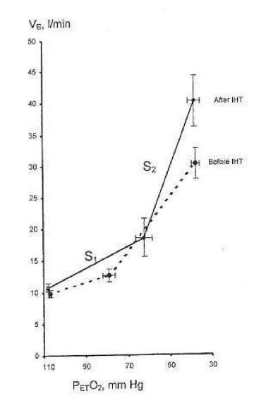
= 150 mmHg) representing central chemoreflex response, and hypoxic (Graphic = 50 mmHg) representing the addition of central and peripheral chemoreflexes responses. The slope of each isoxic response represents sensitivity of the chemoreflex to CO2. The inflection point at which ventilation starts to increase in response to increasing Graphic is the ventilatory recruitment threshold (VRT), where the chemoreflex neural drive to breathe exceeds a drive threshold and starts to produce an increase in pulmonary ventilation. Ventilation below VRT represents non-chemoreflex drives to breathe and is known as the basal ventilation. The differences in ventilation between isoxic rebreathing lines at any given isocapnic Graphic can be used to calculate the hypoxic ventilatory response (indicated by vertical arrows). Note that the choice of isocapnic Graphic affects the magnitude of the measured HVR even within the same subject (Duffin, 2007), with higher HVRs measured at higher isocapnic Graphicvalues in the illustrated example. Note also that the magnitude of HVR provides little information about the characteristics of the control of breathing model, as HVR magnitude is dependent on the combination of central and peripheral chemoreflex responses.

There was no difference in the non-chemoreflex drives to breathe between highlanders and lowlanders, as indicated by similar basal (below VRT) ventilations in the two populations (Table 2).

The highlanders had decreased VRTs compared to lowlanders during both hypoxic and hyperoxic rebreathing tests. The leftward shift of the VRTs in highlanders suggests that a lower Graphic was required to exceed the VRT in highlanders compared to lowlanders. Since both central and peripheral chemoreceptors are actually [H+] sensors, interpretation of this result should consider the acid–base status in both populations. According to the Henderson–Hasselbach equation (Nunn, 1993), the relationship between [H+] and Graphiccan be described as follows:Formulawhere [HCO3] is the bicarbonate ion concentration. In a hypothetical sea-level resident at sea-level, [H+] is approximately 40 nM l−1, Graphic is 40 mmHg and [HCO3] is 24 mM l−1. At altitude, hypoxia-induced hyperventilation results in a reduction of Graphic that leads to a reduction in [H+] and respiratory alkalosis according to the above equation. Highlanders compensate for respiratory alkalosis by presumably reducing their [HCO3] through increased renal excretion, thereby restoring the Graphic/[HCO3] ratio to sea-level values and normalizing [H+].

For example, if hypoxia-induced hyperventilation reduced CO2 from 40 to 30 mmHg, and HCO3− fell from 24 to 18 mM l−1, then the overall ratio of CO2/HCO3− would be maintained at 5/3 as in sea-level lowlanders, but normal [H+] of 40 nM l−1 would be achieved at a lower Graphic of 30 mmHg rather than 40 mmHg, as at sea-level. Considering that the highlanders in our study have an adapted acid–base status (Santolaya 1989), the observed difference in VRTs can be explained by the altered [H+]–Graphic relationship in highlanders compared to lowlanders, with the assumption that the chemoreceptor thresholds [H+] are similar (Duffin, 2005).

The sensitivity of the central chemoreceptor to CO2, as indicated by the ventilatory sensitivity during hyperoxic rebreathing, was lower in highlanders compared to lowlanders (2.5 ± 0.4 vs. 4.2 ± 0.3 l min−1 mmHg−1, P = 0.011). Hyperoxia effectively silences the peripheral chemoreceptor (Lloyd & Cunningham, 1963; Mohan & Duffin, 1997), and therefore the ventilatory sensitivity measured during hyperoxic rebreathing can be taken as a measure of central chemoreceptor sensitivity (Duffin, 2007).

Since the ventilatory response to hypercapnia decreases with age (Nishimura et al. 1991; Jones et al. 1993; Poulin et al.1993; McGurk et al. 1995) and increases with weight (Marcus et al. 1994), the observed lower central CO2 chemosensivity in our highlander subjects may be the result of their older age and smaller body size.

Ventilatory response to hypoxia in highlanders was markedly different from that in lowlanders. Unlike lowlanders, who responded to hypoxia by increasing the sensitivity of their ventilatory response to CO2 (Mohan & Duffin, 1997), the highlanders seemed to decrease their VRT in response to hypoxia with no change in the sensitivity to CO2.

The ventilatory response to hypoxia in sea-level residents is mediated by peripheral chemoreceptors via an increase in the peripheral chemoreceptor sensitivity to [H+] (Cunningham, 1987; Torrance, 1996; Kumar & Bin-Jaliah, 2007). A lack of increase in ventilatory sensitivity to CO2 with induction of hypoxia in highlanders suggests that their peripheral chemoreceptors are relatively insensitive to CO2.

Other possible mechanisms of CO2-independent peripheral responses to hypoxia may include a hypoxia-induced increase in the carotid body tonic drive to breathe, changes in systemic hormonal mediators, an altered cerebral spinal fluid-buffering capacity at the central chemoreceptor or an alteration in cerebral vascular reactivity leading to a higher [H+] at central chemoreceptor.

Specifically, we showed that Himalayan highlanders have decreased central and absent peripheral chemoreceptor sensitivity to CO2, and that they are sensitive to hypoxia, albeit via a different mechanism than that observed in lowlanders at sea-level. A blunted central and an absent peripheral ventilatory sensitivity to CO2 in Himalayan highlanders may stabilize their ventilatory controller by reducing the overall gain in the feedback part of the controller circuit, thereby reducing altitude-related breathing instability (Ainslie & Duffin, 2009)

The non-chemoreflex drives to breathe were similar between Himalayan highlanders and sea-level lowlanders.

Ukjent sin avatar

Etiology of Myofascial Trigger Points

Om triggerpunkter og hvordan muskelspenninger gir oksygenmangel som så fører til melkesyreopphopning. Nevner også at capillærer trekker seg sammen. En annen studie som det refereres til viser bilder og grafer av hvordan dette skjer; økt blodoppsamling pga trange kapillære vener der blodet strømmer ut fra triggerpunktet.

http://www.ncbi.nlm.nih.gov/pmc/articles/PMC3440564/#__ffn_sectitle

«Since the capillary blood pressure ranges from approximately 35 mm Hg at the beginning (arterial side) to 15 mm Hg at the end of the capillary beds (venous side), the capillary blood flow is temporarily obstructed during muscle contractions. The blood flow recovers immediately with relaxation, which is consistent with its normal physiological mechanism. In dynamic rhythmic contractions, intramuscular blood flow is enhanced by this contraction-relaxation rhythm, also known as the muscular pump. During sustained muscle contractions, however, muscle metabolism is highly dependent upon oxygen and glucose, which are in short supply.»

«Since oxygen and glucose are required for the synthesis of adenosine triphosphate (ATP), which provides the energy needed for muscle contractions, sustained contractions may cause a local energy crisis due to the lack of oxygen. To guarantee an adequate supply of ATP, the muscle can switch within a few seconds to anaerobic glycolysis. »

«Under anaerobic circumstances, however, most of the pyruvic acid produced during glycolysis is converted into lactic acid, thereby increasing the intramuscular acidity (pH). Most of the lactic acid diffuses out of the muscle into the bloodstream; post-exercise lactic acid is washed out within 30 minutes after exercise. Unfortunately, when the capillary circulation is restricted, as in sustained low-level contractions, this process comes to a standstill.»

«Small increases of the H+ concentration, as seen with inflammation, heavy muscle work, and ischemia, are sufficient to excite muscle group IV endings, contributing to mechanical hyperalgesia and central sensitization (15).»

«They identified 2 contributing factors, namely an increase in the volume of the vascular compartment, and an increased outflow resistance. Increased outflow resistance could be due to muscle contractures at the TrP that compress the capillary or venous bed. Sustained low-level contractions are common in the workplace where many occupations rely on prolonged postures, as seen in musicians, supermarket cashiers, computer operators, hairdressers, and dentists, among others.» Bilder ref til denne studies: http://www.ncbi.nlm.nih.gov/pmc/articles/PMC3493167/

«Hägg suggested that the continuous activity of these motor units in sustained contractions causes overuse muscle fiber damage, especially to the Type I fibers during low-level activities, which he summarized in his Cinderella hypothesis21. It is conceivable that in sustained low-level contractions and in dynamic repetitive contractions, ischemia, hypoxia and insufficient ATP synthesis in type I motor unit fibers are responsible for increasing acidity, Ca2+ accumulation, and subsequently sarcomere contracture. Furthermore, starting with the sarcomere (super-) contraction, the intramuscular perfusion slows down and ischemia and hypoxia will occur. This may lead to the release of several sensitizing substances causing peripheral sensitization15,22

«A key factor is the local ischemia, which leads to a lowered pH and a subsequent release of several inflammatory mediators in muscle tissue. Hocking proposed an interesting counterargument, which deserves further exploration. Whether overuse mechanisms are the crucial initiating factor or persistent nociceptive input remains a point of debate and further study.»

Forklaring på hva Hocking mener:
«Rather than looking at overuse mechanisms, Hocking maintains that persistent nociceptive input causes the formation of TrPs through central sensitization of the C-fiber nociceptive withdrawal reflex and plateau depolarization of withdrawal agonist alpha-motor neurons and compensatory reticulospinal motor facilitation of antigravity muscles and plateau depolarization of withdrawal antagonist alpha-motor neurons (33). «

Ukjent sin avatar

Hypoxia-generated superoxide induces the development of the adhesion phenotype

Viktig studie om mekanismen bak hvordan hypoxi gir arrvev (adhesions) i kroppen. Relatert til hyperventilering vil lite CO2 gir hypoxi og sammen med trange blodkar vil de utsatte stedene i kroppen utvikle arrvev mellom muskler og nerver. Nevner hvordan antioksidanter er viktig for å unngå arrvev, spesielt etter operasjoner. Og motsatt, at oksidanter kan skape arrvev fra friskt vev. Nevner også hvordan nitratreaksjoner er med å skaper arrvev, så mulig at CO2 bidrar med å dempe nitratreaksjonene og dermed dempe dannelsen av arrvev. Den viser også at det kan være mulig å få arrvev celler om til å bli normale celler.

http://www.ncbi.nlm.nih.gov/pmc/articles/PMC2574925/

«Adhesion fibroblasts exhibit higher TGF-β1 and type I collagen expression as compared to normal peritoneal broblasts. Furthermore, exposure of normal peritoneal fibroblasts to hypoxia results in an irreversible increase in TGF-β1 and type I collagen. We postulated that the mechanism by which hypoxia induced the adhesion phenotype is through the production of superoxide either directly or through the formation of peroxynitrite. »

«Hypoxia treatment resulted in a time-dependent increase in TGF-β1 and type I collagen mRNA levels in both normal peritoneal and adhesion fibroblasts.»

«In contrast, treatment with SOD, to scavenge endogenous superoxide, resulted in a decrease in TGF-β1 and type I collagen expression in adhesion fibroblasts to levels seen in normal peritoneal fibroblasts; no effect on the expression of these molecules was seen in normal peritoneal fibroblasts. »

«In conclusion, hypoxia, through the production of superoxide, causes normal peritoneal fibroblasts to acquire the adhesion phenotype. Scavenging superoxide, even in the presence of hypoxia, prevented the development of the adhesion phenotype. These findings further support the central role of free radicals in the development of adhesions.»

«Postoperative adhesions are a significant source of impaired organ functioning, decreased fertility, bowel obstruction, difficult reoperation, and possibly pain (1,2)

The following popper user interface control may not be accessible. Tab to the next button to revert the control to an accessible version.

Destroy user interface c»

«The processes that result in either normal peritoneal tissue repair or the development of adhesions include the migration, proliferation, and/or differentiation of several cell types, among them inflammatory, immune, mesothelial, and fibroblast cells (3)

«Hypoxia, resulting from tissue injury, has been suggested to play an important role in wound healing, and may therefore be a critical factor in the development of postoperative adhesions (4,7)

The following popper user interface control may not be accessible. Tab to the next button to revert the control to an accessible version.

Destroy user interface contrHypoxia is known to trigger the expression of TGF-β1, which consequently increases the expression of extracellular matrix proteins, including type I collagen (4) 

The following popper user interface control may not be accessible. Tab to the next button to revert the control to an accessible version.

Destroy user interface contr«Type I collagen synthesis has been shown to be crucially dependent on the availability of molecular oxygen in tissue culture, animal, and human wound healing experiments (8,9)

The following popper user interface control may not be accessible. Tab to the next button to revert the control to an accessible version.

Destroy user interface controlMoreover, exposure of normal peritoneal fibroblasts to hypoxia irreversibly induces TGF-β1 and type I collagen to levels seen in adhesion fibroblasts (4,10)

«Additionally, hypoxia is known to acutely promote superoxide (O2.−) generation from disparate intracellular sources that include xanthine dehydrogenase oxidase (11), mitochondrial electron transport chain (12), and NAD[P]H oxidase (13).

In biological systems, superoxide dismutase (SOD) protects against the deleterious actions of this radical by catalyzing its dismutation to hydrogen peroxide plus oxygen, (14) Whereas SOD breaks down O2.−, xanthine oxidase synthesizes O2.−. Xanthine oxidase appears to be one of the major superoxide-producing enzymes (14)«

«Scavenging superoxide restores both TGF-β1 and type I collagen mRNA levels in adhesion fibroblasts to levels observed in normal peritoneal fibroblasts»
«Normal peritoneal and adhesion fibroblasts treated with super-oxide dismutase, a O2.− scavenging enzyme, exhibited a dose–response decrease (0, 5, 10, 15, and 20 units/ml) in TGF-β1 and type I collagen mRNA levels in adhesion fibroblasts while not effecting normal peritoneal fibroblasts (Figs. 3A and B).»

«Scavenging superoxide during hypoxia exposure protects against the development of the adhesion phenotype»

«Peroxynitrite treatment increased the adhesion phenotype markers, TGF-β1 and type I collagen»

«Adhesion fibroblasts are myofibroblasts, defined as transiently activated fibroblasts exhibiting features intermediate between those of smooth muscle cells and fibroblasts, including the expression of α-SM actin (29,21) and a depleted antioxidant system (22). In normal wound healing, as the wound resolves, the cellularity decreases and the myofibroblasts disappear by apoptosis (23). However, in several pathological cases, including fibrosis, myofibroblastic differentiation persists and causes excessive scarring (24,25)

«This is further supported by the fact that when O2.− was scavenged, there was in a significant decrease in TGF-β1 and type I collagen in adhesion fibroblasts to levels seen in normal peritoneal fibroblasts. »

«Reactive oxygen species (ROS) are involved in TGF-β-stimulated collagen production in murine embryo fibroblasts (NIH3T3), and the effect of glutathione depletion on TGF-β-stimulated collagen production may be mediated by facilitating ROS signaling (37)

«Reactive oxygen and nitrogen intermediates control the synthesis of cytokines and growth factors in several in vitro models (40). For instance, they modulate the expression and/or release of monocyte chemoattractant protein-1 (41,42), tumor necrosis factor-α, interleukin (IL)-1 (43,44), IL-8 (45,46), platelet-derived growth factor (47,48), and TGF-β1 (49). «

«Adhesion fibroblasts exhibited a significantly lower level of nitric oxide (NO) and higher protein nitration as compared to normal peritoneal fibroblasts, although there was no difference in the iNOS expression level between the two cell lines (17,50,51). This strongly indicates that adhesion fibroblasts use NO to form ONOO−, and consequently their basal ONOO− levels are higher than normal peritoneal fibroblasts. «

«Thus, treatment with SOD might affect the homeostasis of myofibroblasts by inducing cell death or the phenotypic reversion of myofibroblasts into normal fibroblasts. »

«Our results clearly indicate that hypoxia generated O2.− is a key player in the formation of the adhesion phenotype. This became evident when normal peritoneal fibroblasts were treated with SOD under hypoxic conditions and no change in adhesion markers was seen.»


«In this model, hypoxia-generated O2.− exerts its effect directly by enhancing the expression of TGF-β1, which consequently leads to elevated levels of type I collagen, a hallmark of the adhesion phenotype.»

Ukjent sin avatar

The underlying mechanisms for development of hypertension in the metabolic syndrome

Studie som nevner «alt» om insulinresistens og hva det gjør i kroppen: magefett, symatisk overstimulering, oksidativt stress og blodkardysfunksjon, renin-angiotensin, betennelser og søvnapne.

http://www.nutritionj.com/content/7/1/10

«Visceral obesity, insulin resistance, oxidative stress, endothelial dysfunction, activated renin-angiotensin system, increased inflammatory mediators, and obstructive sleep apnea have been proposed to be possible factors to develop hypertension in the metabolic syndrome. These factors may induce sympathetic overactivity, vasoconstriction, increased intravascular fluid, and decreased vasodilatation, leading to development of hypertension in the metabolic syndrome.»

«As shown in Figure 1, accumulated visceral adipose tissue produce and secrete a number of adipocytokines, such as leptin, tumor necrosis factor-α (TNF-α), interleukin-6 (IL-6), angiotensinogen, and non-esterified fatty acids (NEFA), which induce development of hypertension [11]. «

«Insulin resistance is the main pathophysiologic feature of the metabolic syndrome. Several mechanisms connect insulin resistance with hypertension in the metabolic syndrome. An anti-natriuretic effect of insulin has been established by accumulating data indicating that insulin stimulates renal sodium re-absorption [1214]. This anti-natriuretic effect is preserved, and may be increased in individuals with insulin resistance, and this effect may play an important role for development of hypertension in the metabolic syndrome [15].»

«NEFA has been reported to raise blood pressure, heart rate, and α1-adrenoceptor vasoreactivity, while reducing baroreflex sensitivity, endothelium-dependent vasodilatation, and vascular compliance[28]. Insulin resistance increases plasma leptin levels, and leptin has been reported to elevate sympathetic nervous activity, suggesting that leptin-dependent sympathetic nervous activation may contribute to an obesity-associated hypertension [29]. Accumulating data suggest that metabolic syndrome is associated with markers of adrenergic overdrive [30].»

«In rats with the metabolic syndrome, induced by chronic consumption of a high fat, high refined sugar [31], hypertension is associated with oxidative stress [32], avid nitric oxide (NO) inactivation, and down-regulation of NO synthase (NOS) isoforms and endothelial NOS activator[32], suggesting that oxidative stress and endothelial dysfunction may be strongly associated with development of hypertension in the metabolic syndrome. Further, recent evidences suggest that oxidative stress, which is elevated in the metabolic syndrome [33], is associated with sodium retention and salt sensitivity [34].»

«The renin-angiotensin system (RAS) plays a crucial role in blood pressure regulation, by affecting renal function and by modulating vascular tone. The activity of the RAS appears to be regulated by food intake, and overfeeding of rodents has been reported to lead to increased formation of angiotensin II in adipocytes [38]. «

«Recent cohort studies have demonstrated that high-sensitivity C-reactive protein (hsCRP) independently presents additive prognostic values at all levels of metabolic syndrome [45]. Ridker PM, et al. suggest a consideration of adding hsCRP as a clinical criterion for metabolic syndrome[45]. Abnormalities in inflammatory mediators have been also reported to be implicated with development of hypertension.»

«TNF-α is involved in the pathophysiology of hypertension in the metabolic syndrome. TNF-α stimulates the production of endothelin-1 and angiotensinogen [48,49].»

«IL-6 stimulates the central nervous system and sympathetic nervous system, which may result in hypertension [54,55]. The administration of IL-6 leads to elevation in heart rate and serum norepinephrine levels in women [56]. Further, IL-6 induces an increase in plasma angiotensinogen and angiotensin II [57], leading to development of hypertension.»

«Patients with OSA are often considered to be obese, however, Kono M et al. reported that OSA was associated with hypertension, dyslipidemia, and hyperglycemia, independent of visceral obesity [59]. «

Ukjent sin avatar

Exercise-induced dehydration with and without environmental heat stress results in increased oxidative stress.

Viser at dehydrering øker oksidativt stress og dermed celleskade i trening.

http://www.ncbi.nlm.nih.gov/pubmed/21980993

«Oxidized glutathione (GSSG) increased significantly postexercise in dehydration trials only»

«Finally, both 90-min and 5-km TT performances were reduced during only the DE-W trial, likely a result of combined cellular stress, hyperthermia, and dehydration. »

Fra fecebook artikkel om studien:

«The results of this study show that regardless of temperature, oxidative stress (as measured by oxidized glutathione in the blood) was increased in exercise-induced dehydration, but this increase did not occur when hydration was normal. »

«This study shows that both heat and dehydration influence cellular mechanisms during exercise, but it is not the oxidative stress per se that reduced exercise performance during to the heat-dehydration trial. «