Ukjent sin avatar

The Anatomy of Dorsal Ramus Nerves and Its Implications in Lower Back Pain

Om hudnære nerver sin rolle i korsryggsmerter. Beskriver spesielt nervene ved L1-2 som går ned til huden i korsryggen og hoftene. Nevner flere interessante diagnosekriterier. Men som de legene og kirurgene forskerne er, har de kun nerveblokkade og kirurgi i som behandlingmuligheter, fullstendig ignorante til mulighetene i manuell behandling. Selv nerveblokkade har 85% av pasientene får mer enn 50% bedring i symptomene, ikke så annerledes enn hva vi forventer med manuell behandling og en intelligent tilnærming til nervesystemet.

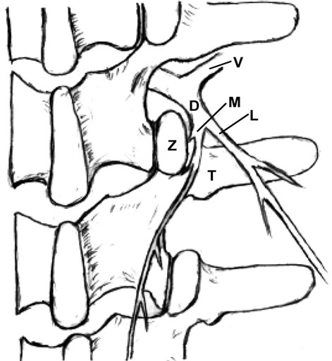
Each spinal dorsal ramus arises from the spinal nerve and then divides into a medial and lateral branch. The medial branch supplies the tissues from the midline to the zygapophysial joint line and innervates two to three adjacent zygapophysial joints and their related soft tissues. The lateral branch innervates the tissues lateral to the zygapophysial joint line.

Clinically, L1 and L2 are the most common sites of dorsal rami involvement.

The etiologies of low back pain are numerous. Anatomically, lumbar muscle strain [1,2], lumbar zygapophysial joint syndrome [3-11], instability of the lumbar spine [12], discogenic back pain [1,13], and sacroiliac joint syndrome [2] can cause low back pain. Mechanical pressure on the nerve roots, which may interfere with venous return of the nerve root [14], epidural fibrosis [15], perineural and intraneural fibrosis [16], are additional factors to consider. Additionally, some authors have suggested that the iliolumbar ligament inserting on the lumbar spine is a source of back pain [17].

Anatomically, Bogduk’s work exposed the medial branches of the lumbar spinal dorsal rami as a potential player in low back pain [23-25]. Sihvonen et al. blocked the medial dorsal ramus branch, which resulted in relief of muscle spasms and they suggested that this treatment would aid in improving lumbopelvic rhythm and reducing low back pain [26-29]. These studies supported spinal dorsal ramus as a potential pain generator.

http://file.scirp.org/Html/9-2400120/b3c5afd3-5aa8-4350-9ecc-22fe3e3cc027.jpg

http://file.scirp.org/Html/9-2400120/c6242d6a-bc68-420a-bd92-3c9e7fbafa2d.jpg

The distribution area of each dorsal ramus is characterized by an overlapping multiple segmental innervations, e.g., the L4 zygapophysial joint is innervated by the L3 and L4 medial branches. Therefore, if single dorsal ramus is irritated proximally, a patient could experience pain at the distal site of this nerve distribution (referred pain). This phenomenon mimics radicular pain, for example, a patient with L4-5 herniated disc experiences pain on his dorsal foot. Thus, a local anesthetic injected to the referred pain area will not relieve pain, because the pain is caused by irritation at the proximal dorsal ramus (see below) [36].

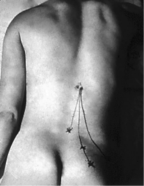
In patients with the spinal dorsal ramus mediated pain, their symptoms usually are on one side and are exacerbated by lumbar extension and/or rotation. This pain may radiate to the ipsilateral low back and buttock region (referred pain) [21,22] (Figure 2). Some patients may present paraspinal muscle spasm (Figure 3(a)). Hyperesthesia may present in the affected dermatome [10,21,22,31,35, 45,46].

http://file.scirp.org/Html/9-2400120/e412d7c3-e859-4500-b82d-396e71f6b638.jpg

The zygapophysial joint line demarcates the distribution of the medial and lateral branches. Pain at between the midline and the zygapophysial joint line or the paraspinal sacroiliac region is caused by on irritated medial branch. Pain lateral to the zygapophysial joint line with radiation to the lateral iliac crest is induced by the lateral branch involvement [19,22]. When the common dorsal ramus is involved, pain will be at the territories of both medial and lateral branches [36,38,47] (Figure 2).

There are some clinical findings in the patient with the spinal dorsal ramus mediated low back pain. The patient usually points to pain at the distal low back (referred pain) [19,22,47]. When the patient bends forward, there is usually a palpable step-off at the affected spinous processes and this is typically two to three segments above the referred pain [47]. There is a palpably widened space and deep tenderness between the spinous processes below the stepoff. With deep palpation of the junction of the same level lateral zygapophysial joint and proximal transverse process, the patient will experience pain and referred pain [19-22,36,47] (Figure 2). Additionally, patients may present an ipsilateral segmental muscle spasm, and a mild scoliosis at the affected vertebral level when the medial branch is involved (Figure 3(a)) [36,47]. If the lateral branch is involved, palpating the longissimus and the iliocostalis muscles can be painful [19-22,35,36,47]. Maigne’s examination techniques are to provoke pain by applying pressure to the lateral aspect of the spinous processes and rubbing the ipislateral facet at the thoracolumbar junction [19-22]. Other findings such as motor, sensory and straight leg raising tests are unremarkable. When the low back pain patient presents pain with radiation below the knee and positive nerve root signs such as loss of sensory or motor function or reflexes in the distribution of the ventral ramus, the ventral ramus involvement (lumbar radiculopathy) should be considered [1].

Any abnormality of the zygapophysial joint such as vertebral malrotation or muscle spasm as well as structural changes of the zygapophysial joint such as subluxation, degeneration, bony proliferation, capsular/ligamentous hypertrophy or fracture can irritate the common dorsal ramus and medial branch, and induce clinical symptoms [18,44,47,48]. Ossification of the mammilloaccessory ligament may cause an entrapment neuropathy and low back pain [18,24,33].

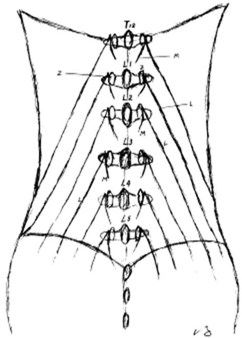
Chen and colleagues [51] dissected the spinal dorsal rami from T12 to the sacrum and conducted biomechanical studies. Their study demonstrated that the L2 dorsal rami bore the greatest stretching force and tensile stress when the specimens were flexed and rotated to the contralateral side.

Spinal dorsal ramus mediated back pain can occur at any level of the human spine [18,21,22,44,52,53]. For low back pain mediated by dorsal ramus, the primary pain is commonly at the thoracolumbar junction [19-22, 38,44]. Within the thoracic region, the coronal orientation of the zygapophysial joints grants spine free rotation. However, this rotation is limited by a rigid rib cage, except at the T10-12 levels because of floating ribs. The upper lumbar facets also have a relative coronal orientation. Therefore, spine rotation is relatively free at the thoracolumbar junction and the greatest shear force occurs at the more mobile upper lumbar segments. This normal spinal movement can cause zygapophysial joint separation or rotation. If these movements occur rapidly or overcome the body’s physiological limit, they can cause stretching tension and irritation to the dorsal ramus, resulting in low back pain [20,40,44,54]. Shao and his colleagues reported that seventy four percent (74%) of the 1263 patients with spinal dorsal ramus mediated low back pain had the pain originating from L1 and/or L2 dorsal ramus [36].

http://file.scirp.org/Html/9-2400120/3fe33e99-0bf8-4fbf-ac3c-867b9de28c6b.jpg

Additionally, Zhou has reported in his retrospective study, that in 41 patients with spinal dorsal ramus mediated lower back pain, after the selective spinal dorsal ramus injection, 84% of these patients received greater than 50% and more than two months of pain reduction [74]. These patients also reported improvement in their daily activities and decrease of their pain medications [74].

Normally, this type of pain originates at L1 or L2 dorsal rami, and the pathogeneses can be multiple factors which irritate the dorsal ramus. The back pain induced by dorsal ramus irritation can occur in the cervical [51] and thoracic spine [50] as well. Therefore, spinal dorsal ramus mediated back pain should be appropriately called “spinal dorsal ramus syndrome (SDRS)”.

The clinical presentations of dorsal ramus mediated back pain and zygapophysial syndrome can be overlapping. However, there are some distinctions. The thoracolumbar junction is the most common site of spinal dorsal ramus mediated back pain [21,22,36], while, zygapophysial joint syndrome commonly occurs at the lower lumbar zygapophysial joints such as L5-S1 and L4-5 [5,6,9,43,57, 58].

Ukjent sin avatar

Stress and the inflammatory response: a review of neurogenic inflammation.

Betennelser kan både skapes og opprettholdes av nervesystemet.

Det er velkjent av betennelser økes av stresshormonet kortisol når vi stresser fordi kortisol senker immunfunksjon og dermed øker betennelsestilstander.

Men denne studien beskriver hvordan stress øker nevrogen betennelse (betennelse i nervesystemet), som kan forklare årsaken til at alt som vanligvis bare er litt ukomfortabelt blåses opp og blir vondere når vi er i langvarig stress.

Man tenker vanligvis på sansenerver som noe som sender signaler fra kroppen, gjennom ryggraden og opp til hjernen. Men molekyler kan faktisk gå andre veien i nervetrådene også. Fra ryggraden og UT i kroppen. Når vi stresser sender nervecellene ut et stoff som kalles Substans P, sammen med andre betennelsesøkende stoffer. Der hvor nervetrådene ender (i ledd, i huden eller i organer) blir det en lokal betennelsesreaksjon som bidrar til smerte. Substans P er spesielt assosisert med smertetilstander.

Forskeren konkluderer også med at dette er en viktig årsak til hvordan kronisk stress kan bidra til kroniske betennelsessykdommer som arterosklreose i blodårene eller betennelser i organene.

Beste måten å roe ned et stresset nervesystem er meditasjon med Autonom pust (5-6 pust i minuttet).

http://www.ncbi.nlm.nih.gov/pubmed/12480495

The subject of neuroinflammation is reviewed. In response to psychological stress or certain physical stressors, an inflammatory process may occur by release of neuropeptides, especially Substance P (SP), or other inflammatory mediators, from sensory nerves and the activation of mast cells or other inflammatory cells.

Central neuropeptides, particularly corticosteroid releasing factor (CRF), and perhaps SP as well, initiate a systemic stress response by activation of neuroendocrinological pathways such as the sympathetic nervous system, hypothalamic pituitary axis, and the renin angiotensin system, with the release of the stress hormones (i.e., catecholamines, corticosteroids, growth hormone, glucagons, and renin). These, together with cytokines induced by stress, initiate the acute phase response (APR) and the induction of acute phase proteins, essential mediators of inflammation. Central nervous system norepinephrine may also induce the APR perhaps by macrophage activation and cytokine release. The increase in lipids with stress may also be a factor in macrophage activation, as may lipopolysaccharide which, I postulate, induces cytokines from hepatic Kupffer cells, subsequent to an enhanced absorption from the gastrointestinal tract during psychologic stress.

The brain may initiate or inhibit the inflammatory process.

The inflammatory response is contained within the psychological stress response which evolved later. Moreover, the same neuropeptides (i.e., CRF and possibly SP as well) mediate both stress and inflammation.

Cytokines evoked by either a stress or inflammatory response may utilize similar somatosensory pathways to signal the brain. Other instances whereby stress may induce inflammatory changes are reviewed.

I postulate that repeated episodes of acute or chronic psychogenic stress may produce chronic inflammatory changes which may result in atherosclerosis in the arteries or chronic inflammatory changes in other organs as well.

Ukjent sin avatar

Respiratory Dysregulation in Anxiety, Functional Cardiac, and Pain Disorders

Svært mye interessant i denne studien om pusten og CO2. Spesielt avsnittene om at kronisk smerte endrer pustemønsteret og senker CO2 nivået i kroppen.

http://www.ncbi.nlm.nih.gov/pubmed/11530714

http://www.mental-mechanics.org/pdf/Anxiety/FH%20Wilhelm%20et%20al%20-%20Respiratory%20dysregulation%20review.pdf

CHRONIC PAIN
Acute pain results in shortness of breath and an increase in ventila- tion (Nishino, Shimoyama, Ide, & Isono, 1999). A commonly used pain provocation in the laboratory is immersion of a limb into almost freezing water (cold pressor test), which is reliably followed by reduc- tions of PetCO2 among healthy people. (On the other hand, partial or full immersion of the face in cold water causes a modest reduction in ventilation, a component of the diving response). Patients who experi- ence intense chronic pain show these respiratory-related changes over extended periods. For example, migraine headache patients were found to have significantly lowered PetCO2 levels during an attack compared to controls and to migraine-free periods (Hannerz & Jogestrand, 1995), and there were even respiratory abnormalities immediately before an attack (Zhao, Sand, & Sjaastad, 1992). Glynn, Lloyd, & Folkhard (1981) examined arterial pH and PCO2 in 52 chronic pain patients (e.g., back pain, cancer-related pain). PCO2 was mark- edly lowered in these patients, and nerve blockade of pain resulted in a significant rise in PCO2.

Interestingly, blood pH was normal, indicat- ing a long-term blood chemistry compensation for chronic hyperven- tilation. In a sleep study of fibromyalgia patients, a high incidence of respiratory abnormalities such as periodic breathing were found, and arterial PCO2 was lowered in a subgroup of patients (Sergi et al., 1999). Many clinicians, including one of the present authors (Gevirtz), have had the opportunity to measure PetCO2 levels in hun- dreds of chronic muscle pain patients, and the clinical impression is that these levels are almost universally low (c.f., Timmons & Ley, 1994). Of course, pain may also play a role in the increased ventilation found in the FCD patients discussed above, especially during acute episodes of chest pain.

The increased ventilation during acute pain is likely a component of the fight-flight response, preparing the individual for immediate action and sometimes for being attacked or maybe injured. Interest- ingly, recent evidence from animal studies indicates that acute hyperventilation has anesthetic effects via the adrenergic and endogenous opiate system (Ide et al., 1994a, 1994b). Thus, the increased ventila- tion that first served to activate an individual for a fight may have the beneficial side effect of relieving pain if the fight is lost.

So far, no study we know of has examined if the chronic hyperventi- lation exhibited by pain patients is of any benefit to their pain experi- ence (and thus a coping strategy), is only a side effect of the intense pain, or makes their pain worse. One would expect that chronic hyper- ventilation is not healthy in these patients, as it is in other clinical groups, because it interferes with blood homeostatic mechanisms and can lead to a variety of physical symptoms. It has been suggested that by numbing pain, hyperventilation may become a short-term adaptive process with long-term negative consequences (Conway, 1994). Inter- esting in this context is that opioids are frequently prescribed to chronic pain patients to suppress their pain, and they typically also suppress ventilation via central nervous pathways, sometimes to a lethal extent. In summary, there is some initial evidence that hyper- ventilation plays a role in chronic pain, and some mediating mecha- nisms have been identified. However, most of the pain-hypocapnia relationship in chronic pain syndromes is not well understood.

Chronic Pain
Slow abdominal breathing is often taught as a relaxation technique in preparation for acute pain, such as surgery or childbirth, and it also helps patients counteract their tendency to hyperventilate during such events. As described above, the chronic hyperventilation that can accompany long-lasting pain may be especially problematic because it may have long-term negative organismic effects. It is therefore logi- cal that breathing training could be a valuable asset in the overall treat- ment of chronic pain disorders. However, no data are currently avail- able on the role of breathing training as a systematic intervention in these disorders. It is one author’s (Gevirtz) clinical experience that breathing training is in fact a powerful tool in a comprehensive pain management protocol. This is also a common assumption of most bodywork therapies of pain (c.f., Clifton-Smith, 1998). Here again, the capnometry readings are used to illustrate the physiological basis of the symptomatology.

Muscular pain can result from chronically tense muscles. Hubbard, Gevirtz, and their colleagues recently showed that a sympathetically mediated pathway to muscle spindles (trigger points), rather than pathways to muscle fibers, plays an important role in the maintenance of chronic muscular pain (Gerwin, Shannon, Hong, Hubbard, & Gevirtz, 1997; Hubbard & Berkoff, 1993; McNulty, Gevirtz, Hub- bard, & Berkoff, 1994). Psychological stress increased the activity of these spindles, which suggests that stress reduction could alleviate chronic muscle pain. Thus, relaxation induced by slow diaphragmatic breathing may have a beneficial effect on the activation of these spin- dles and reduce general muscle tension.

Ukjent sin avatar

Huden – vårt største og viktigste sanseorgan

huden

ALLE (!!!) muskel- og ledd terapeuter tar først og fremst på huden. Og ALLE gjør alt de kan for å overse det. Vi kan trygt si at huden er undervurdert i behandling av menneskekroppen. Spesielt siden ALLE behandlingseffekter egentlig er sekundæreffekter av det som skjer i hudens nervesystem og hjernens opplevelse av dette.

Huden er vårt største organ. Og vårt mest sensitive organ. Det er så tettpakket av sensoriske nervetråder at vi kan si den er «utsiden av hjernen». Vi er faktisk så godt beskyttet av huden at det er kun hjernens opplevelse av det som skjer med huden som avgjør hva som skjer med vevet under huden. Alle som tar på huden i en behandlingssituasjon er fullstendig underlagt nervesystemets reaksjon på berøringen.

Muskler, ledd og bindevev gjør bare det nervesystemet befaler. Når vi vet hvilken direkte kobling huden har til nervesystemet og hjernen kan vi også rette behandlingskonseptene våre direkte på det som faktisk gir behandlingseffekt: Nervesystemet

Når vi trykker på et triggerpunkt, et ømt område av en muskel, så finnes det svært få trykksensitive nerver i selve muskelen. De finnes hovedsaklig  i huden og vevet rett under huden. Smerten vi opplever av trykket kommer altså ikke av at vi trykker på muskelen, men at vi trykker på huden. Selv om det kjennes ut som at vi trykker hardt og dypt inn vevet, så er det hudens nerver som reagerer på trykket. Om vi bedøver hudens nerver når vi f.eks. er støle, så forsvinner trykksensitiviteten også. Når vi jobber med å dempe smerte trenger vi altså ikke trykke hardt inn i muskelen, vi trenger bare å ha en litt smartere tilnærming til huden.

Smerte er en subjektiv opplevelse som er ment å beskytte kroppen. Det innebærer et tett samarbeid mellom hjernens registrering av faresignaler fra huden og dens interne kart over kroppen. Fra et evolusjonært perspektiv er det større farer i omgivelsene enn internt i kroppen. Derfor dekker de fare-registrerende nervene (nociceptorer) hele kroppens overflate. Det er også noe av grunnen til at vi føler oss trygge og slapper bedre av når noen tar på huden vår på en behagelig og ikke-truende måte.

Det krever en ganske stor omveltning for å innse hudens rolle i smertebehandling. Det er veldig mye som vi tidligere har tatt for gitt som må snus på hodet. Vi må lære oss å legge merke til noe medisinsk vitenskap har brukt 200 år på å overse.

Med en helt ny forståelse av menneskekroppen og behandling av nervesystemet kan vi også behandle smertetilstander direkte, men uten å gi ny smerte slik de «gamle» behandlingsformene gjør. De som overser huden, vårt største og viktigste sanseorgan, og (håpløst) prøver å trykke fingrene igjennom den.

Med den nye behandlingsformen DermoNeuroModulation snur vi alt på hodet og behandler smerte UTEN å gi ny smerte. Når vi vet hvordan huden og nervesystemet fungerer trenger vi kun å gi en behagelig og interaktiv behandling av huden, og smerte dempes umiddelbart.

Ukjent sin avatar

The Pathogenesis of Muscle Pain

Viktig studie fra S.Mense som nevner mange aspekter av muskelsmerter, som trykksensitivitet, sentral sensitering og referert smerte.

http://www.cfids-cab.org/cfs-inform/Neuroendocrin/mense03.pdf

The typical muscle nociceptor responds to noxious local pressure and injections of BKN; however, in animal experiments, receptors also can be found that are activated by one type of noxious stimulation (mechanical or chemical) only. This finding indicates that different types of nociceptors are present in skeletal muscle, similar to the skin in which mechano-, mechano-heat-, and poly-modal nociceptors have been reported to exist [12••].

The sensitization is associated with a decrease in the mechanical threshold of the receptor so that it responds to weak pressure stimuli. The sensitized muscle receptor still is connected to nociceptive central nervous neurons and thus elicits subjective pain when weak mechanical stimuli act on the muscle. This sensitization of muscle nociceptors is the best established peripheral mechanism explaining local tenderness and pain on movement of a pathologically altered muscle.

Of these neuropeptides, SP is of particular interest because, in experiments on fibers from the skin, SP has been shown to be predominantly present in nociceptive fibers [26]. The peptides are released during excitation of the ending and influence the chemical milieu of the tissue around the receptor. This means that a nociceptor is not a passive sensor for tissue-threatening stimuli, but actively changes the micromilieu in its vicinity by releasing neuropeptides. SP has a strong vasodilating and permeability increasing action on small blood vessels.

Mechanism of referral of muscle pain

The expansion of the input region of the inflamed GS muscle nerve likely underlies the spread and referral that is common in patients with muscle pain. The mechanisms mentioned previously can explain referral as follows: when a muscle is damaged, the patient will perceive local pain at the site of the lesion. If the nociceptive input from the muscle is strong or long-lasting, central sensitization in the dorsal horn neurons is induced, which opens silent synapses and leads to an expansion of the target area of that muscle in the spinal cord (or brain stem). As soon as the expansion reaches sensory neurons that supply peripheral areas other than the damaged muscle, the patient will feel pain in that area outside the initial pain site. In the area of pain referral, no nociceptor is active and the tissue is normal. The referral is simply caused by the excitation induced by the original pain source, which spreads in the central nervous system and excites neurons that supply the body region in which the referred pain is felt. This way, a trigger point in the temporalis muscle can induce pain in the teeth of the maxilla when the trigger point-induced central excitation spreads to sensory neurons that supply the teeth [43•].

Ukjent sin avatar

Muscle Pain: Mechanisms and Clinical Significance

En studie til fra Siegfried Mense, om muskelsmerter. Han har ikke fått med seg at trykksensitive nerver kun finnes i huden. Og han har misforstått litt i forskjellene mellom hud-smerter og muskel-smerter siden han sier at hud-smerter ikke kan ha utstrålende effekt. Han har tydeligvis ikke ikke inkludert subcutane nerver i sin vurdering.

Men mye interessant i denne studien likevel. Spesielt vektleggingen av at lav pH er den viktigste bidragsyteren til muskelsmerter.

Han nevner at input fra muskel-nociceptorer har større relevans i ryggmargen enn input fra huden. Derfor er betennelser og lav pH de viktigste drivkreftene i kroniske smerter.

Nevner også at smerter henger sammen, f.eks. at trapezius kan stramme seg for å beskytte brachialis, slik at smerten kjennes i trapezius, mens problemet egentlig sitter i brachialis.

Beskriver også triggerpunkter, men sier at det foreløpig er veldig mange ubesvarte spørsmål om denne teorien.

http://www.ncbi.nlm.nih.gov/pmc/articles/PMC2696782

Muscle pain is a major medical problem: in, the majority (60% to 85%) of the population has had (nonspecific) back pain of muscular origin at some time or other (lifetime prevalence) (1). Pain evoked by myofascial trigger points has a point prevalence of approximately 30% (2). More than 7% of all women aged 70 to 80 years suffer from the fibromyalgia syndrome (e1). In an Italian study, musculoskeletal pain was found to be the most common reason that patients consulted a doctor (3). Thus, treating physicians should be aware of the mechanisms of muscle pain, insofar as they are currently understood.

Subjective differences between muscle pain and cutaneous pain

Muscle pain Cutaneous pain
Electrical nerve stimulation induces only one pain Electrical nerve stimulation induces a first pain and a second pain
Poorly localizable Well-localized
Tearing, cramping, pressing quality Stabbing, burning, cutting quality
Marked tendency toward referral of pain No tendency toward referral of pain
Affective aspect: difficult to tolerate Affective aspect: easier to tolerate

Muscle pain is produced by the activation of specific receptors (so-called nociceptors): these receptors are specialized for the detection of stimuli that are objectively capable of damaging tissue and that are subjectively perceived as painful. They consist of free nerve endings and are connected to the central nervous system (CNS) by way of unmyelinated (group IV) or thinly myelinated (group III) fibers. They can be sensitized and activated by strong mechanical stimuli, such as trauma or mechanical overloading, as well as by endogenous inflammatory mediators including bradykinin (BK), serotonin, and prostaglandin E2 (PGE2).

Two activating chemical substances are particularly important for the generation of muscle pain: adenosine triphosphate (ATP) and protons (H+ ions).

ATP activates muscle nociceptors mainly by binding to the P2X3 receptor molecule, H+ mainly by binding to the receptor molecules TRPV1 (transient receptor potential vanilloid 1) and ASICs (acid-sensing ion channels) (4).

ATP is found in all cells of the body and is released whenever bodily tissues of any type are injured.

A drop in pH is probably one of the main activators of peripheral nociceptors, as many painful disturbances of muscle are associated with low pH in muscle tissue.

Nerve growth factor (NGF) also has a connection to muscle pain: NGF is synthesized in muscle and activates muscle nociceptors (e2). NGF synthesis is increased when a muscle is inflamed (e3).

Acidic tissue pH is one of the main activating factors leading to muscle pain. Practically all pathological and pathophysiological changes of skeletal muscle are accompanied by a drop in pH, among them

  • chronic ischemic states,
  • tonic contractions or spasms,
  • myofascial trigger points,
  • (occupationally induced) postural abnormalities, and
  • myositides.

The neuropeptides stored in muscle nociceptors are released not only when peripheral stimuli activate the nerve endings, but also when spinal nerves are compressed. In this type of neuropathic pain, action potentials are generated at the site of compression and spread not only centripetally, i.e., toward the central nervous system, but also centrifugally, i.e., toward the nociceptive endings, where they induce the release of vasoactive neuropeptides. In this way, neurogenic inflammation comes about, characterized by hyperemia, edema, and the release of inflammatory mediators (8). The inflammatory mediators sensitize the muscle nociceptors and thereby increase neuropathic pain.

The sensitization of the muscle nociceptors by endogenous mediators such as BK and PGE2 is one of the reasons why patients with muscle lesions suffer from tenderness to pressure on the muscle, and from pain on movement or exercise. It is also the reason why many types of muscle pain respond well to the administration of non-steroidal anti-inflammatory drugs (NSAID), which block prostaglandin synthesis.

An influx of nervous impulses from muscle nociceptors into the spinal cord increases the excitability of posterior horn neurons to a greater extent than one from cutaneous nociceptors (9).

Two main mechanisms underlie the overexcitability of spinal nociceptive neurons:

A structural change of ion channels, rendering them more permeable to Na+ and Ca2+, is the short-term result of an influx of nociceptive impulses into the spinal cord. Among other effects, this causes originally ineffective («silent» or «dormant») synapses to become effective.

A change of gene transcription in the neuronal nucleus, leading to a modification of synthetic processes, causes new ion channels to be synthesized and incorporated into the nerve cell membrane. The long-term result of central sensitization is a nociceptive cell whose membrane contains a higher density of ion channels that are also more permeable to ions. This explains the hyperexcitability of the cell. Glial cells, too, particularly microglia, can contribute to the sensitization of central neurons by secreting substances such as tumor necrosis factor a (TNF-a) (8).

The increased excitability of spinal neurons and the spread of excitation within the CNS are the first steps in the process of chronification of muscle pain. The endpoint of chronification consists of structural remodeling processes in the CNS that open up new pathways for nociceptive information and cause pain to persist over the long term. Patients with chronic muscle pain are difficult to treat, because the functional and structural changes in the CNS need time to regress. The fact that not all muscle pain becomes chronic implies that chronification requires not only the mechanisms just discussed, but also other ones, e.g., a genetic predisposition.

Pain arising in muscle is more likely to be referred pain than pain arising in the skin. Referred pain is pain that is felt not (only) at its site of origin, but at another site some distance away. A possible mechanism of referred pain is the spread, within the spinal cord, of excitation due to the muscle lesion (9) (figures 2 and ​and3).3). As soon as the excitation reaches sensory posterior horn neurons that innervate an area beyond the site of the original muscle lesion, the patient feels referred pain in that area, even though none of the nociceptors in it are activated (13).


An example is shown in figure 3: a stimulus delivered to the myofascial trigger point (MTrP) in the soleus muscle causes only mild local pain, while the patient feels more severe (referred) pain in the sacroiliac joint. No conclusive answers are yet available to the questions of why muscle pain is more likely than cutaneous pain to be referred, why it is usually not referred to both proximal and distal sites, and why pain referral is often discontinuous. There is, however, a well-known discontinuity of spinal topography between the C4 and T2 dermatomes.

The main reason why pain arises in muscle spasm is muscle ischemia, which leads to a drop in pH and the release of pain-producing substances such as bradykinin, ATP, and H+.

The vicious-circle concept of muscle spasm – muscle pain causes spasm, which causes more pain, etc. – should now be considered obsolete. Most studies have shown that muscle pain lowers the excitability of the α-motor neurons innervating the painful muscle (14) (a «pain adaptation» model) (15).

Muscle spasm can be precipitated by, among other things, pain in another muscle. Thus, a spasm-like increase EMG activity in the trapezius muscle has been described in response to painful stimulation of the biceps brachii muscle (16). Another source of muscle spasms is pathological changes in a neighboring joint. These sources of pain must be deliberately sought.

In a widespread hypothesis on the origin of MTrP’s (19), it is supposed that a muscular lesion damages the neuromuscular endplate so that it secretes an excessive amount of acetylcholine. The ensuing depolarization of the muscle cell membrane produces a contraction knot that compresses the neighboring capillaries, causing local ischemia. Ischemia, in turn, leads to the release of substances into the tissue that sensitize nociceptors, accounting for the tenderness of MTrP’s to pressure. Substances of this type have been found to be present within the MTrP’s of these patients (20). This supposed mechanism leaves many questions unanswered but is currently the only comprehensive hypothesis on the origin of MTrP’s.

Patients with MTrP’s often have pain in three locations:

  • at the site of the MTrP itself,
  • at the origin or insertion of the affected muscle, because of pulling by the muscle fibers that have been stretched by the contraction knots,
  • and referred pain outside the MTrP (figure 3).

Because the MTrP is cut off from its blood supply by compression of the local microcirculation, oral NSAID’s are not very effective against TrP pain.

Ukjent sin avatar

Functional Anatomy of Muscle – Muscle, Nociceptors and Afferent Fibers

Svært mye interessant om nervesystemets rolle i muskler og smerte.

Spesielt at det er ingen frie nerveender i muskelcellene, men bare i blodkarene i musklene. Derfor reagerer vi med smerte på betennelser og lav pH i blodet, mens trykksensitiviteten kun sitter i huden.

Den nevner at pH sensibiliteten er den viktigste smertebidraget fordi pH synker i de fleste patologiske tilstander, f.eks. hard trening eller skade.

Den nevner at det er mer SP (Substans P, som er relatert til smerte) i huden enn i muskler.

Nevner at frie nerveender ikke går til kapillærer eller muskelceller, bare til arterioler og venuler.

Nevner også innervering av bindevev, og at dette feltet foreløpig er lite studert og oversett. Spesielt viser de til at Toracolumbar Facia (i korsryggen) har størst innervasjon av C-fiber nociceptorer(som inneholder SP) under huden, og litt i multifidene.

En nociceptor er ikke bare en passiv mottaker av impulser, men er også en aktiv deltaker i vevets tilstand når det gjelder betennelser og blodsirkulasjon for de sender nevropetider ut fra doresalhornet til vevet (antidromiske impulser). Altså motsatt vei av reseptor-signalretningen.

CGRP virker vasodilerende, mens SP gjør at blodkarveggenes permeabilitet øker. Når permeabiliteten øker siver det ut proteiner og stoffer som egentlig ikke skal være i vevet, og da økes betennelser og immunsystemets aktivitet. Så det er SP vi ønsker å dempe først og fremst.

http://www.google.com/url?sa=t&rct=j&q=&esrc=s&source=web&cd=1&cad=rja&ved=0CCsQFjAA&url=http%3A%2F%2Fwww.springer.com%2Fcda%2Fcontent%2Fdocument%2Fcda_downloaddocument%2F9783540850205-c1.pdf%3FSGWID%3D0-0-45-935848-p173845615&ei=_GJEUqyUOevn4QSBl4HQDg&usg=AFQjCNEpyWrolHywvUNyw_Cwa8yWTiEUnw&sig2=OJLZ3XrnTalCUUqE-uhCeQ&bvm=bv.53217764,d.bGE

The predominant location of free nerve endings supplied by group IV fibers was the adventitia of arterioles and venules. Surprisingly, muscle fibers themselves did not receive direct innervation by free nerve endings. Group III afferents generated not only free nerve endings but also paciniform corpuscles, whereas group IV fibers terminated exclusively in free nerve endings.The high sensitivity of the free nerve endings to chemical stimuli, particularly to those accompanying inflammatory lesions or disturbances of the microcirculation, may be related to their location on or in the walls of the blood vessels. The finding that the muscle fibers proper are not supplied by free nerve endings (Reinert et al. 1998) may relate to the clinical experience that muscle cell death is usually not painful, at least not if it occurs slowly, as for instance during muscular dystrophy, polymyositis, or dermatomyositis. A different situation is tearing of a muscle fiber bundle, which can be extremely painful. In this condi- tion, many muscle cells are destroyed simultaneously and release their contents (e.g., K+ ions and ATP) in the interstitial space, from where they can diffuse to the next nociceptive endings.
In skeletal muscle, the free nerve endings appear to be distributed quite evenly in the proximodistal direction. At least, in a quantitative evaluation of the inner- vation density by neuropeptide-(SP- and CGRP-) containing fibers, no difference between the proximal and distal portions of the rat gastrocnemius–soleus (GS) muscle was found (Reinert et al. 1998). Therefore, a higher innervation density at the transition zone between muscle and tendon is not a probable explanation for the frequent pain in this region.

However, in the same study the nerve fiber density in the peritendineum (the connective tissue around a tendon) of the rat calcaneal tendon was found to be several times higher than that in the GS muscle. In contrast, the collagen fiber bundles of the tendon tissue proper were almost free of free nerve endings. The high fiber density in the peritendineum may explain the high prevalence of tenderness or pain in the tissue around the tendon and the insertion site. The scarcity of nerve endings in the center of the tendon may relate to the clinical observation that (incomplete) ruptures of the tendon may occur without pain.

Judging from their respon- siveness to pain-producing agents, the following receptor molecules are likely to be relevant for muscle pain and tenderness (Mense and Meyer 1985; Caterina and David 1999; McCleskey and Gold 1999; Mense 2007):

  • Bradykinin (BKN) receptors (B1 and B2). BKN is cleaved from blood plasma proteins when a blood vessel breaks or increases its permeability so that plasma proteins enter the interstitial space. In intact tissue, BKN excites nerve endings by the activation of the BKN receptor molecule B2, whereas under pathological conditions (e.g., inflammation) the receptor B1 is the predominant one (Perkins and Kelly 1993; for a review of receptor molecules mediating the effects of classic inflammatory (pain-producing or algesic) substances, see Kumazawa 1996).
  • Serotonin receptors (particularly 5-HT3). Serotonin (5-hydroxytryptamin, 5-HT) is released from blood platelets during blood clotting. The stimulating effects of serotonin on nociceptive terminals in the body periphery are predomi- nantly mediated by the 5-HT3 receptor (at present, more than 15 different 5-HT receptors are known in the CNS). The serotonin concentrations released in the tissue are usually not sufficient to excite nociceptors directly, but they can sen- sitize them, i.e., make them more sensitive to other pain-producing agents such as BKN.
  • Prostaglandins, particularly prostaglandin E2 (PGE2). Prostaglandins (PGs) are released in a pathologically altered muscle by the enzymatic action of cycloox- igenases. PGE2 binds to a G protein-coupled prostanoid receptor (EP2) in the membrane of the nociceptive ending. Similarly to serotonin, PGE2 sensitizes nociceptors rather than exciting them under (patho)physiological circumstances (Mense 1981).
  • Acid-sensing ion channels (ASICs). ASICs constitute a family of receptor molecules that are sensitive to a drop in pH and open at various pH values. The channel proteins react already to small pH changes, for instance from pH 7.4 to 7.1. This receptor family (for instance ASIC1 and ASIC3) is particularly impor- tant for muscle pain, because almost all pathologic changes in muscle are accom- panied by a drop in tissue pH, e.g., exhausting exercise, ischemia, and inflammation (Immke and McCleskey 2003). In these conditions, the pH of the muscle tissue can drop to 5–6. The proton-sensitive nociceptors may also be of importance for the induction of chronic muscle pain. Repeated intramuscular injections of acidic solutions have been reported to induce a long-lasting hyper- algesia (Sluka et al. 2001).
  • P2X3 receptors. This receptor is a subtype of the purinergic receptors that are activated by ATP and its derivatives (Burnstock 2007; Ding et al. 2000). ATP is the energy-carrying molecule in all cells of the body; accordingly, it is present in every tissue cell. It is released from all tissues during trauma and other pathologic changes that are associated with cell death. For this reason, ATP has been considered a general signal substance for tissue trauma and pain (Cook and McCleskey 2002). ATP is particularly important for muscle pain, because it is present in muscle cells in high concentration (Stewart et al. 1994). When injected into human muscle, ATP causes pain (Mo ̈rk et al. 2003).
  • Transient receptor potential receptor subtype 1 (TRPV1) formerly called VR1. This receptor is one of the most important molecules for the induction of pain. The natural stimulant for the TRPV1 receptor is Capsaicin, the active ingredient of chilli peppers (Caterina and Julius 2001). The receptor is also sensitive to an increase in H+-concentration and to heat, with a threshold of approximately 39C. Its endoge- nous ligands are H+-ions.
  • Other TRP receptors. TRPV4 is a mechanosensitive ion channel that is sensitive to both weak and strong (noxious) intensities of local pressure (Liedtke 2005). It may be the receptor for mediating pain evoked by pinching and squeezing.
  • Tyrosine kinase A (TrkA) receptor. The ligand of this receptor is NGF (Caterina and David 1999). NGF is well-known for its sensitizing action on nociceptors in the body periphery and neurons in the CNS; it is synthesized in muscle, and its synthesis is increased during pathophysiological changes of the muscle (e.g., inflammation, Menetrey et al. 2000; Pezet and McMahon 2006).
  • Glutamate receptors. There is evidence indicating that the NMDA receptor (one of the receptors for glutamate) is present on nociceptive endings in masticatory muscles. Injections of glutamate into the masseter muscle in human subjects induced a reduction in pressure pain threshold which was attenuated by coinjection with ketamine, an NMDA receptor antagonist (Cairns et al. 2006).
Substances exciting muscle nociceptors independent of membrane receptors.
  • Hypertonic saline: injections of NaCl solutions (4.5–6.0%) have long been and still are used to elicit pain from deep somatic tissues (Kellgren 1938; for review, see Graven-Nielsen 2006).
  • Potassium ions: The most likely explanation for the excitatory action of high concentrations of extracellular potassium ions is a depolarization of the membrane potential due to a reduction of the inside–outside potassium gradient (usually the potassium concentration inside the axon is much higher).

DRG cells projecting in a cutaneous nerve have been reported to contain SP, CGRP, and somatostatin (SOM).

In comparison to skin nerves, muscle nerves appear to contain less SP. This finding makes sense, because the vasodilatation and plasma extrava- sation caused by the release of SP and CGRP from free nerve endings (see below) would be dangerous for skeletal muscles, since many of them are surrounded by a tight fascia. Therefore, an SP-induced muscle edema would result in a high increase in interstitial pressure, and could cause muscle necrosis.

In a study on functionally identified DRG cells employing a combination of electrophysiological and immunohistochemical techniques, Lawson et al.(1997) reported that cells terminating in cutaneous nociceptive endings showed a strong tendency to express SP, particularly if they had a slow conduction velocity or a small soma in the DRG. 

The peptides are synthesized in the somas of the DRG or in ganglion cells of cranial nerves. They are transported to both the central and the peripheral terminal of the primary afferent unit.

In a quantitative evaluation of neuropeptide-containing free nerve endings and preterminal axons (both characterized by varicosities) in the GS muscle of the rat, most endings were found around small blood vessels (arterioles or venules), whereas capillaries and the muscle cells proper were not contacted by these end- ings.

Efferent or motor fibers conduct action potentials from the CNS to the periphery; their soma is located in the spinal cord or brainstem and the fibers leave the CNS via the ventral root or cranial nerve motor roots. An exception are postganglionic sympathetic fibers whose cell bodies are mostly located in the sympathetic trunk (e.g., vasomotor fibers that constrict blood vessels).

The nerve to a locomotor muscle in the cat (the lateral GS) is composed of approximately one-third myelinated (720) and two-thirds unmyelinated (2,480) fibers (Table 2.2; Mitchell and Schmidt 1983; Stacey 1969). Nearly one quarter of the myelinated (group III) fibers had nociceptive properties in neurophysio- logical experiments (Mense and Meyer 1985). Of the unmyelinated fibers, 50% are sensory (group IV), and of these, approximately 55% have been found to be nociceptive in the rat (Hoheisel et al. 2005).

Data obtained from one muscle nerve cannot be transferred directly to other muscle nerves, because considerable differences exist between different muscles. For instance, neck muscle nerves of the cat contain unusually high numbers of sensory group III receptors (Abrahams et al. 1984). One possible explanation for these differences is that the muscles have different functions and environmental conditions: in contrast to the neck muscles, which must register the orientation of the head in relation to the body in fine detail, the locomotor hindlimb muscles often have to contract with maximal strength and under ischemic conditions.

In addition to nociceptors, there are other muscle receptors whose function is essential for the understanding of muscle pain:

  • Muscle spindles are complex receptive structures that consist of several specialized muscle fibers (the so-called intrafusal muscle fibers; the name is derived from their location inside the spindle-shaped connective tissue sheath. Accordingly, all the “normal” muscle fibers outside the spindle are “extrafusal” fibers). Muscle spindles measure the length and the rate of length changes of the muscle, i.e., their discharge rate increases with increasing muscle length and with increasing velocity of the length change.
  • Golgi (tendon) organs measure the tension of the muscle. They are arranged in series with the extrafusal muscle fibers; their location is the transition zone between muscle and tendon. The supplying fiber is the Ib afferent, whose structure is identical to the Ia fiber (thick myelin sheath and high conduction velocity). The receptor has a much simpler structure than the muscle spindle; it consists of receptive endings that are interwoven between the collagen fiber bundles of the tendon.
  • Muscle spindles and Golgi organs are proprioceptors, i.e., they measure the internal state of the body.
  • Pacinian corpuscles (PC) and paciniform corpuscles. These receptors do not respond to static pressure; they require dynamically changing mechanical stimuli, and are best excited by vibrations of relatively high frequency (close to 300 Hz; Kandel et al. 2000). The receptive ending is formed like a rod, and covered by several concentric membranes which give the receptor an onion-like appearance in cross-sections.

At present, little information is available about the innervation of fascia. This is an important gap in our knowledge, because fascia is an important component of the musculoskeletal system and likely to contribute to many forms of pain that are subsumed under the label “muscle” pain. One example is low back pain: The thoracolumbar fascia (TF) plays an essential role in body posture and trunk move- ments (Bogduk and Macintosh 1984). It is not only a passive transmitter of mechanical forces of the low back and abdominal muscles but also contractile by itself (Schleip et al. 2005).

In the connective tissue around the superficial lamina of the TF we found many CGRP- and SP-containing free nerve endings. The majority of the fibers were located in the subcutaneous layer, as well as between the fascia and the surface of the multifidus muscle (Fig. 2.8). The SP-positive endings are of particular interest, because they are thought to be nociceptors.
The loose connective tissue around the TF is probably deformed during any trunk movement, and therefore the free nerve endings are strategically situated to sense any disorders in these movements. It is conceivable that overload of the fascia puts mechanical stress and irritation on the endings, and thus may contribute to low back pain.
SP then releases histamine from mast cells, and together with CGRP these agents cause vasodilatation and an increase in vascular permeability of the blood vessels around the active ending. The result is a shift of blood plasma from the intravascular to the interstitial space. Outside the blood vessel, BKN is cleaved from the plasma protein kallidin, serotonin (5-HT) is set free from platelets, and PGs (particularly PGE2) from endothelial and other tissue cells. All these substances sensitize nociceptors. Thus, the main tissue alteration induced by a nondestructive noxious mechanical stimulus is a localized region of vasodilatation, edema, and sensitized nociceptors.
A nociceptor is not a passive sensor of tissue-threatening stimuli; it actively influences the microcirculation and chemical composition of the intersti- tial space around it.
If a noxious stimulus activates only one part of the ending, the action potentials originating in that region of the ending can invade antidromically (against the normal direction of propagation) those branches of the ending that were not excited by the stimulus. These antidromic action potentials release neuropeptides from the unstimulated branches. The whole process is called the axon reflex.  It is assumed to be the reason for the visible wheal and flare around a cutaneous lesion.

The vascular permeability is increased mainly by SP (and by the neurokinins A and B; Gamse and Saria 1985), whereas CGRP is assumed to act primarily as a vasodilator. There is evidence showing that CGRP enhances the plasma extravasation induced by SP and neurokinins A and B, but reduces the vasodilatory action of SP by desensitizing muscle arterioles to the peptide (O ̈ hle ́n et al. 1988).

The area of wheal and flare after a localized damage to the skin – for instance around a needle prick – could be an indicator of the extent of the excited nocicep- tive ending.

The size of the receptive fields (RFs) of cutaneous polymodal nociceptors was found to be less than 2 mm in cat (Bessou and Perl 1969) and 6–32 mm in rabbit (Kenins 1988). A receptive field is that region of the body from which a receptive ending (or a central sensory neuron) can be excited. The above figures are larger than the reported length of the branches of a nociceptor ending (a few hundred mm; Stacey 1969).

The release of SP, CGRP, neurokinin A, and other agents from nociceptors is the central factor in the cascade of events that lead to neurogenic inflammation in the periphery (Lembeck and Holzer 1979). Neurogenic inflammation is characterized by tissue edema and infiltration by immune cells, i.e., it exhibits the major histo- logical signs of a (sterile) inflammation. It develops whenever action potentials are generated not at the receptive ending but somewhere along the course of primary afferent units (spinal nerve or dorsal root). The action potentials propagate both to the CNS (causing pain) and to the peripheral ending (causing release of neuropep- tides and neurogenic inflammation). The published data indicate that vasodilatation can be elicited by antidromic stimulation of both Ad- and C fibers, but increase in vascular permeability and plasma extravasation by stimulation of C fibers only.

Neuropathies and radiculopathies and other pathological conditions that are asso- ciated with antidromic activity in sensory nerve fibers are examples of such events (Marchand et al. 2005). Neurogenic inflammation is likely to increase the dysesthe- sia and pain of patients suffering from neuropathies.

Inflammatory disorders are usually accompanied by sensitization of peri- pheral nociceptors, which is one source of inflammatory pain (for details, see Chap. 3). 

Ukjent sin avatar

Ny behandlingsform mot hodepine og nakkespenninger på Verkstedet

Muskelspenninger helt øverst i nakken bidrar til mange problemer. F.eks. hodepine, spenningsmigrene, kjevespenning, nakkeplager, bevegelsessmerter i nakken, dårlig søvn, m.m.. Spesielt smerter i panna og tinningene har ofte utgangspunkt i området øverst i nakken.

På Verkstedet har vi nå en ny behandlingsform som løser opp dette problemområdet på en svært behagelig og overraskende effektiv måte, og med en eksepsjonelt elegant forklaringsmodell som er på vei til å revolusjonere forståelsen av smertebehandling verden over.

I området som kalles Occiput helt øverst i nakken har vi mange muskler som styrer hodets balanse i alle vinkler. Muskel- og leddterapeuter(fysio, kiro, osteo, massører, osv) tenker vanligvis at det er muskelspenningene som er problemet og at det gjør vondt fordi musklene er stive og inneholder triggerpunkter, eller fordi et ledd er låst. Men med denne nye behandlingsformen innser vi at det er nervesystemet som har problemer, ikke musklene eller leddene. Musklene og leddene gjør bare det nervesystemet befaler. Og det er nervesystemet helt ytterst i huden som reagerer på trykk, IKKE muskelene eller bindevevet. Når vi trykker på en muskel eller et triggerpunkt så er det altså ikke trykket på muskelen du kjenner, men trykket på nervene helt ytterst i huden.

Når vi endelig innser at det er de sensoriske nervene rett under huden som reagerer på trykk og opplevelsen av smerte eller stråling ut panna og tinningene, så kan vi også behandle disse direkte.

I konvensjonell medisin har man skjønt at det er nervetrådene som har problemer. Når det har blitt et seriøst problem kaller de det Trigeminusnevralgi eller Occipetal Nevralgi. Men her behandles disse nervene med f.eks. nedfrysing, bedøvelse, Botox eller avbrenning. I forhold til den nye behandlingformen vi har tatt inn på Verkstedet er dette unødvendig smertefulle og inngripende behandlingsmodeller. Og det værste av alt, de er dyre og har ikke spesielt god effekt heller.

Forskere har også sett at om man bedøver huden når man er støl etter trening, så forsvinner smerten. Selv når man er støl, hvor det kjennes ut som at man har vondt inni muskelen og det er vondt å bevege muskelen, så er det egentlig i nervene helt ytterst i huden som bidrar til smerteopplevelsen. Dette er ikke lett å forstå fordi det er ikke det vi har lært, og det er ikke slik det kjennes ut. Vi har lært at muskelsmerter sitter i musklene, og vi kan verifisere det ved at det «kjennes ut» som at det sitter i musklene. Men som forskerne her har påvist, smerten opprettholdes egentlig i nervetrådene i huden.  https://mariusblomstervik.no/2013/07/14/c-tactile-fibers-contribute-to-cutaneous-allodynia-after-eccentric-exercise/

Med vår nye behandlingsmetode får du en umiddelbar release av muskelspenninger og smerte øverst i nakken. Ikke fordi vi masserer hardt, bedøver eller brenner av nerver, men fordi vi gir huden en behaglig og mild strekk som åpner opp for de minste nervetrådene i huden. De får mer blodsirkulasjon, mer plass, mer næring, og en behagelig stimuli som demper smerte i løpet av noen få minutter.

I forskning regner man at en smertereduksjon på 2 poeng i en 10-poeng skala er «statistisk signifikant». Klienter rappoerterer en umiddelbar reduksjon på 6-8 poeng med denne behandlingsformen. Det er ganske radikalt.

Og det er svært overraskende at noe så enkelt og noe så behagelig kan gi en så stor endring i smerteopplevelsen. Selv de som er vandt til behandling hvor «vondt skal vondt fordrive» lar seg overraske av effekten i dette behandlingskonseptet.

Behandlingsformen kalles DermoNeuroModulation. Legg merke til dette navnet. Dette er begynnelsen på en revolusjon i behandling av smertetilstander. Dermo betyr huden, Neuro betyr nerver, Modulation betyr å endre. Altså, vi endrer nervesystemets respons med behandling av hudens nerver.

Varigheten av forbedringen er avhengig av mange faktorer, bla. ernæring, trening, stressreduksjon og alvorlighetsgraden man kommer med i utgangspunktet. For de fleste gir det en radikal bedring allerede etter første gang, og vanligvis trenger man 2-4 ganger for å få de mest solide resultatene. Noen trenger mer, noen trenger mindre, men ALLE blir overrasket over effekten.

Ta kontakt om du ønsker å se om denne behandlingsformen kan hjelpe for din hodepine eller nakkespenninger. Trykk her for online bestilling 

Ukjent sin avatar

Stop Chasing Pain… It’s Not In The Body, It’s In The Brain!

Reception – Protection – Integration – Modulation – Discrimination

Dette skjer i nervesystemet:

1. sanseceller registrerer huden/omverdenen og det interne miljøet, balanse, sanser, osv.

2. signalene går videre igjennom reptilhjernens amygdala og hippokampus, som kobler det til tidligere opplevelser og erfaringer og avgjør om vi skal inn i en fight-or-flight/freeze modus. Avgjør om vi er i en trussel-situasjon og aktiveres som en refleks.

3. videre opp til cerebellum, lillehjernen, hvor signalene plasseres i hjernens «kart» over kroppen og den forhold til omgivelsene, som resten av hjernen kan bruke til å avgjøre hva den skal gjøre.

4. Derifra reagerer det autonome nervesystem for å modulere effekten basert på hva hjernen velger å gjøre. Sympaticus stimulerer, parasympaticus beroliger.

5. Og deretter kan frontallappen utføre sin diskriminering og bevisste valg som kan tilby nye reaksjonsmønstre til reptilhjernen. I trusselsituasjoner får ikke frontallappen sjangsen til å gjøre jobben sin. Reaksjonene skjer før frontallappen rekker å vurdere situasjonen. Skal man lære nye reaksjonsmønstre må man oppleve reduksjon i trusselsituasjonen først.