Ukjent sin avatar

Recognition of central sensitization in patients with musculoskeletal pain: Application of pain neurophysiology in manual therapy practice

Nevner det aller meste om sentral sensitering og hvordan de gjenkjennes i klinisk praksis.

http://www.thblack.com/links/RSD/ManTher2010_15_135_CSinMusculoskelPain-PainNeurophys.pdf

«Even with acute pain the nervous system undergoes some changes. When tissue is damaged and pain persists for a few days with adaptation of unimodal nociceptors, the responsiveness of polymodal nociceptive endings is enhanced by substances released from various sources (i.e. serotonin released by platelets) (Purves et al., 1997). This process is called primary hyperalgesia or peripheral sensitization of nociceptors, and represents a protective action by the human body in order to prevent further use of damaged structures and consequent further damage of the traumatized and surrounding tissues.»

«Secondary hyperalgesia refers to increased responsiveness of dorsal horn neurons localized in the spinal segments of the primary source of nociception.»

«Central sensitization is defined as an augmentation of responsiveness of central neurons to input from unimodal and polymodal receptors (Meyer et al., 1995). Central sensitization encompasses altered sensory processing in the brain (Staud et al., 2007), malfunctioning of descending anti-nociceptive mechanisms (Meeus et al., 2008), increased activity of pain facilitatory path- ways, temporal summation of second pain or wind-up (Meeus and Nijs, 2007; Staud et al., 2007), and long-term potentiation of neuronal synapses in the anterior cingulate cortex (Zhuo, 2007).»

«The presence of central sensitization in patients with musculoskeletal pain implies an increased complexity of the clinical picture (i.e. an increase in unrelated symptoms and hence a more difficult clinical reasoning process) (Nijs et al., 2009), as well as decreased odds for a favorable rehabilitation outcome (Jull et al., 2007).»

«Central sensitivity syndromes is a term first used by Yunus in 2000 to describe a group of overlapping conditions bound by a common pathophysiological mechanism of central sensitization (Yunus, 2007a).»

«Another example is chronic non-specific low back pain. Some studies provided evidence in support of the presence of central sensitization in patients with non-specific chronic low back pain (Giesecke et al., 2004; Schmidt-Wilcke et al., 2006), while others refute such an association (Hoffman et al., 2005; Julien et al., 2005). It is concluded that central sensitization is present in some cases of chronic non-specific low back pain, possibly representing one of the subgroups of this frequent musculoskeletal disorder (Wand and O’Connell, 2008).»

«The myofascial variety within the heterogeneous group of temporomandibular disorders is also characterized by central sensitization (Yunus, 2007a). Likewise, regional chronic pain conditions that present with tender and/or trigger points in the absence of structural pathology (frequently referred to as myofascial pain syndrome) should alert the manual therapist to the possibility that central sensitization is determining the clinical picture (Yunus, 2007a). However, to our knowledge available evidence in support of central sensitization in patients with myofascial pain syndrome is limited to chronic whiplash associated disorders, temporoman- dibular disorders and chronic non-specific low back pain.»

«Furthermore, various subgroups of headache, chronic tension-type headache (Langemark et al., 1993; Pielsticker et al., 2005) and migraine (Burnstein et al., 2000; Weissman-Fogel et al., 2003) can be viewed as central sensitivity syndromes. Finally, rheumatoid arthritis and osteoarthritis are examples of local musculoskeletal disorders possibly causing continuous activation of polymodal nociceptors that initiate or sustain central sensitization (Yunus, 2007a).»

table1

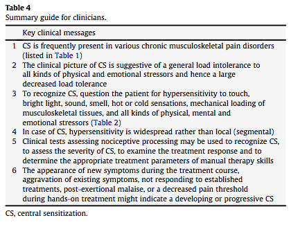
«Central sensitization entails much more than generalized hypersensitivity to pain: it is characterized by an increased responsiveness to a variety of stimuli including mechanical pressure (Desmeules et al., 2004), chemical substances (Morris et al., 1997), cold temperature (Kasch et al., 2005), heat temperature (Meeus et al., 2008), electrical stimuli (Banic et al., 2004; Desmeules et al., 2004), stress, emotions, and mental load. The clinical picture is suggestive of a general intolerance to all kinds of physical and emotional stressors and hence a large decreased load tolerance of the human body in general.»

table2

«An ongoing source of peripheral nociception is required before the process of peripheral sensitization can establish central sensitization (Nijs and Van Houdenhove, 2009). Tissue injury healing and focal pain recovery should occur as soon as possible to prevent development of central sensitization (Vierck, 2006).»

«One of the main characteristics of central sensitization in patients with musculoskeletal pain is a generalized rather than a localized decrease in their pressure pain threshold. Here, ‘generalized’ implies more than a segmental spreading of the symptom area, in that it means that the increased sensitivity is localized at sites segmentally unrelated to the primary source of nociception (e.g. the lower limbs in case of a whiplash trauma).»

«Lower pressure pain thresholds at symptomatic areas most often represent primary hyperalgesia due to sensitized polymodal noci- ceptors within injured musculoskeletal structures. By measuring pressure pain thresholds outside the area of primary nociception, widespread hyperalgesia or secondary hyperalgesia can be detec- ted.»

«In cases of secondary hyperalgesia, a reduced pressure pain threshold in the various tissues innervated by the same segment (or two neighboring segments) can be detected.»

«Findings of numerous areas of hyperalgesia at sites outside and remote from the symp- tomatic site, together with a non-segmental general decrease in pressure pain threshold, may imply a generalized hyperexcitability of central nociceptive pathways (Sterling et al., 2004).»

«Pressure algometry provides a reliable and valid measure of the pressure pain threshold (Vanderweeen et al., 1996; Farasyn and Meeusen, 2003). In the absence of a pressure algometer, manual palpation can be used. Even when a manual therapist is not sus- pecting central sensitization, the finding of generalized hypersen- sitivity to manual palpation during routine clinical examination should alert the clinician.»

«Like every other tissue in the human body, peripheral nerves and nervous tissues (including connective tissue) themselves can become hypersensitive to mechanical stimuli such as tension and pressure.»

«Besides the passive tests listed above (Table 3), altered sensory processing can be demonstrated during exercise. Pain thresholds increase during physical activity in healthy individuals and can stay augmented for up to 30 min post-exercise. This is the result of endogenous opioid release (Koltyn and Arbogast, 1998) and related activation of several (supra)spinal anti-nociceptive mechanisms such as the adrenergic and serotonergic pathways (Millan, 2002).»


«Stress (particularly when chronic) may well trigger lower pain thresholds. This was demonstrated by Suarez-Roca et al. (2008) who reported reduced GABA neurotransmission and consequent hyperalgesia in rats after repeated forced swimming stress.»

«A constant or decreased pain threshold during and following exercise suggests malfunc- tioning of these anti-nociceptive mechanisms (Whiteside et al., 2004) and hence central sensitization. An abnormal pain threshold response to exercise should be regarded as one of the many possible signs of central sensitization.»

table4


Ukjent sin avatar

Systemic inflammatory responses to maximal versus submaximal lengthening contractions of the elbow flexors


Nevner hvordan trening øker betennelse i både spesifikt muskelvev og systemisk, men konkluderer med at det er liten forskjell mellom treningsintensiteter. Forskjellige betennelsesfaktorer. IL-6 er spesielt viktig som anti-inflammatorisk faktor under trening. Kan gi en indikator på måling av stølhet etter trening, siden den øker drastisk etter 3-4 dager som vist i bildet.

http://www.medizin.uni-tuebingen.de/transfusionsmedizin/institut/eir/content/2006/72/article.pdf

«Lengthening muscle contractions elicit an inflammatory response. Within skeletal muscle, pro-inflammatory cytokines are produced, and phagocytic cells invade damaged muscle tissue (for review see ref. (22).»
» Within the systemic circu- lation, the complement cascade is activated, leukocytes are mobilized, and cytokine concentrations increase (22). These inflammatory responses share some similarities with the responses to trauma injury (8).»

«The inflammatory response to contraction-induced injury may be propor- tional to the severity of muscle damage, which in turn is dependent on mechanical loading of muscle during exercise. This relationship is important because the magnitude of the inflammatory response regulates adaptation to muscle injury (32).»

«Furthermore, muscle damage after lengthening contractions (as indicated by loss of muscular strength) corre- lates with blood leukocyte counts and serum C-reactive protein concentration (21).»

«Interleukin (IL)-6 plays an important role in mediating inflammatory responses to exercise. IL-6 is believed to exert anti-inflammatory effects during exercise by inhibiting the production of the pro-inflammatory cytokine tumor necrosis factor (TNF)-α, and stimulating the synthesis of other anti-inflammatory cytokines such as IL-1 receptor antagonist (IL-1ra), IL-10 and soluble TNF-α receptor 1 (sTNF-αR1) (25). IL-6 is produced within, and released from skeletal muscle during exercise in response to glycogen depletion, calcium signaling, changes in blood glucose availability, and to a lesser extent sympathetic activation (9, 11, 13).»

trening og betennelse

Viser at det ikke er noen forskjell på betennelsesfaktorer mellom forskjellige treningsintensiteter:
«The aim of this study was to compare systemic inflammatory responses to sub- maximal versus maximal lengthening contractions. Our data indicate that there were no significant differences in systemic markers of inflammation, despite evidence of greater muscle damage (as indicated by impaired muscular strength) following the maximal versus submaximal contractions.»

Nevner hvordan immunforsvaret reagerer på treningen:
«Elevated blood neutrophil counts are a consistent finding after lengthening contractions of the elbow flexors (17, 27-29). Neutrophils are most likely mobi- lized from endothelial surfaces into the circulation in response to tissue injury such as exercise-induced muscle damage. Once in the circulation, neutrophils travel to the site of injury, and then bind and break down damaged tissue frag- ments. In our study, neutrophil numbers were highest 3 h after exercise.»

«The small increase in serum IL-6 concentration after the submaximal contractions could represent the release of IL-6 from muscle in response to muscle glycogen depletion (13).»

«In conclusion, the present data indicate that although maximal lengthening contractions of the elbow flexors may result in greater muscle damage, this is not accompanied by a greater systemic inflammatory response.»

«Furthermore, although we could not detect differences in systemic inflammation, differences could exist locally within skeletal muscle.»

Ukjent sin avatar

Exercise-induced dehydration with and without environmental heat stress results in increased oxidative stress.

Viser at dehydrering øker oksidativt stress og dermed celleskade i trening.

http://www.ncbi.nlm.nih.gov/pubmed/21980993

«Oxidized glutathione (GSSG) increased significantly postexercise in dehydration trials only»

«Finally, both 90-min and 5-km TT performances were reduced during only the DE-W trial, likely a result of combined cellular stress, hyperthermia, and dehydration. »

Fra fecebook artikkel om studien:

«The results of this study show that regardless of temperature, oxidative stress (as measured by oxidized glutathione in the blood) was increased in exercise-induced dehydration, but this increase did not occur when hydration was normal. »

«This study shows that both heat and dehydration influence cellular mechanisms during exercise, but it is not the oxidative stress per se that reduced exercise performance during to the heat-dehydration trial. «

Ukjent sin avatar

The exercise-induced growth hormone response in athletes.

Studie som gir oversikt over mange faktorer rundt veksthormonøkning i trening.

http://www.ncbi.nlm.nih.gov/pubmed/12797841

«A number of physiological stimuli can initiate hGH secretion, the most powerful, non-pharmacological of which are sleep and exercise. »

«The exercise-induced growth hormone response (EIGR) is well recognised and although the exact mechanisms remain elusive, a number of candidates have been implicated. These include neural input, direct stimulation by catecholamines, lactate and or nitric oxide, and changes in acid-base balance. Of these, the best candidates appear to be afferent stimulation, nitric oxide and lactate. Resistance training results in a significant EIGR.»

«An exercise intensity above lactate threshold and for a minimum of 10 minutes appears to elicit the greatest stimulus to the secretion of hGH. Exercise training above the lactate threshold may amplify the pulsatile release of hGH at rest, increasing 24-hour hGH secretion.»

«In a great many cases, the impact of some of the deleterious effects of ageing could be reduced if exercise focused on promoting the EIGR. «

Ukjent sin avatar

Growth hormone release during acute and chronic aerobic and resistance exercise: recent findings.

Studie som bekrefter hvordan veksthormon påvirkes av trening. Viser at det er et lineært forhold mellom trening og veksthormon. Flere treningsøkter om dagen gir mest økning i veksthormon.

http://www.ncbi.nlm.nih.gov/pubmed/12457419

«Contrary to previous suggestions that exercise-induced GH release requires that a «threshold» intensity be attained, recent research from our laboratory has shown that regardless of age or gender, there is a linear relationship between the magnitude of the acute increase in GH release and exercise intensity. »

» As a result, 24-hour integrated GH concentrations are not usually elevated by a single bout of exercise. However, repeated bouts of aerobic exercise within a 24-hour period result in increased 24-hour integrated GH concentrations.»

«While exercise interventions may not restore GH secretion to levels observed in young, healthy individuals, exercise is a robust stimulus of GH secretion. «

Ukjent sin avatar

Factors contributing to the variability in muscle ageing

Nevner at aldring og muskelatrofi skjer mye på grunn av «low grade inflammation».

http://www.ncbi.nlm.nih.gov/m/pubmed/22902240

«Where an individual cannot change much in his or her genetic constitution, circulating hormones and systemic inflammation, (s)he can still significantly slow the rate of muscle ageing by an adequate dietary intake and regular physical activity. Finally, it is suggested that age-related alterations in the capillary bed may negatively affect muscle mass.»

Ukjent sin avatar

Physical activity, by enhancing parasympathetic tone and activating the cholinergic anti-inflammatory pathway, is a therapeutic strategy to restrain chronic inflammation and prevent many chronic diseases.

Viser hvordan trening demper betennelser ved å aktivere parasympaticus.

http://www.ncbi.nlm.nih.gov/m/pubmed/23395411

«We hypothesize that the «cholinergic anti-inflammatory pathway» (CAP) mediates the anti-inflammatory phenotype and range of health benefits associated with physical activity. The CAP is an endogenous, physiological mechanism by which acetylcholine from the vagus nerve, interacts with the innate immune system to modulate and restrain the inflammatory cascade.»

«Importantly, higher levels of physical activity are associated with enhanced parasympathetic (vagal) tone and lower levels of C-reactive protein, a marker of low-grade inflammation. «

Ukjent sin avatar

Carbon dioxide pressure-concentration relationship in arterial and mixed venous blood during exercise

Om hvordan CO2 endrer seg under trening og at CO2 handler mest om å regulere pH.

http://jap.physiology.org/content/90/5/1798.long

«Although the mean mixed venous PCO 2 rose from 47 Torr at rest to 59 Torr at the lactic acidosis threshold (LAT) and further to 78 Torr at Max, the Cv̄CO2 rose from 22.8 mM at rest to 25.5 mM at LAT but then fell to 23.9 mM at Max.»

«We conclude that changes in buffer base and pH dominate the PCO 2-CCO 2 relationship during exercise, with changes in Hb and blood oxyhemoglobin saturation exerting much less influence.»

«This study discloses several important findings. 

1) During exercise, CCO 2 and [HCO Formula ] do not consistently increase in proportion to PCO 2

2) Because of the acidemia caused by increased lactate production, Cv̄CO2 and mixed venous [HCO Formula ] decrease to near resting values as maximalV˙O 2 is approached, despite increasing Pv̄CO2

3) Above LAT, while Pv̄CO2 increases to high levels, PaCO2 decreases because of ventilatory compensation for the exercise lactic acidosis; consequently, CaCO2 decreases to a greater degree than does Cv̄CO2

4) The increase in Cv̄CO2-CaCO2during exercise is mainly due to the increase in Cv̄CO2 below LAT and the decrease in CaCO2 above LAT.

5) Changes in SO 2 and Hb have minor influences on the PCO 2-CCO 2 relationship during exercise, whereas changes in pH due to changes in buffer base have a major influence.

6) Because pHdecreases more than pHa, there are large errors in calculated Cv̄CO2-CaCO2 when the pH change is ignored. 

7) At rest and during all levels of exercise, over threefourths of the total CO2exchange from the blood to lung gas (i.e., Cv̄CO2-CaCO2) is due to dissociation of [HCO Formula ], whereas less than one-fourth is due to the combination of venoarterial differences in [CO2] and [NH-CO2] at rest to Max.»

«It is clear that the dissociation of [HCO Formula ] plays the dominant role in CO2 exchange at the lung, whereas [CO2] and [NH-CO2] play smaller roles in total CO2 exchange. Although PCO 2differences account for the transfer of CO2 out of blood, >75% of the quantity transferred comes from the dissociation of mixed venous [HCO Formula ].»

«We have shown that Cv̄CO2 in blood actually decreases during exercise above the LAT, despite increasing PCO 2. The major reason for this is that the CO2 dissociation curve is shifted downward when lactic acid is generated during exercise.»

Her er CO2 Disossiation Curve: 

Ukjent sin avatar

Physiological mechanisms dissociating pulmonary CO2 and O2 exchange dynamics during exercise in humans

Beskriver hvordan forholdet mellom CO2 og O2 endrer seg unde trening, og spesielt over melkesyreterskel hvor bikarbonat også begynner å spille inn i homeosasen av pH i blod.

http://ep.physoc.org/content/92/2/347.long

«During incremental exercise, the increased rate of Graphic relative to pulmonary O2 uptake (Graphic) can be used to quantify θL validly if aerobic and hyperventilatory sources can be ruled out, i.e. θL is then attributable to the decrease in muscle and blood [HCO3]. In many cases, however, very rapid incrementation of work rate and/or prior depletion of CO2 stores (by volitional or anticipatory hyperventilation) can yield a ‘false positive’ non-invasive estimation of θL(‘pseudo-threshold’) resulting from a slowing of the rate of wash-in of transient CO2stores.»

«Since the tissue capacitance for CO2 is appreciably greater than for O2 (Farhi & Rahn, 1955), this means that the respiratory exchange ratio (R), i.e. the ratio of the volumes of CO2and O2 exchanged across the tissue of interest per unit time, will differ from that of the respiratory quotient (RQ), i.e. the ratio of the amounts of metabolic CO2 and O2 produced and consumed, respectively, by the tissue per unit time not only across the lung, where it is most typically determined and from which inferences are most typically drawn, but also across the muscle vascular bed itself. »

«But, in addition, skeletal muscle contraction results in a transient metabolic alkalosis in the force-generating units (Steinhagen et al. 1976; Kemp, 2005) and in the venous effluent of the exercising muscle (Wasserman et al. 1997) as a result of the net proton (H+) trapping associated with the high-energy phosphate utilization, i.e. H+ release as ATP is split, and H+ uptake consequent to phosphocreatine (PCr) splitting (Kushmerick, 1997).»

«This transient alkalosis therefore results in a component of the metabolically produced CO2 being retained within the muscle.»

Ukjent sin avatar

Relationship between Hyperventilation and Excessive CO2 Output during Recovery from Repeated Cycling Sprints

Nevner at CO2 ikke er årsak til hyperventillering under trening, men at det er melkesyre. Pusten øker for å fjerne CO2 så syreoverskuddet holdes i balanse.

http://eprints.lib.hokudai.ac.jp/dspace/bitstream/2115/51990/1/repeat-ex-PR.pdf

«During incremental exercise, blood lactate is progressively increased above the VT. This is buffered by the bicarbonate system. This results in progressive reduction of blood bicarbonate ion (Beaver et al. 1986a) and metabolic acidosis. In order to improve this metabolic acidosis, ventilation is driven and becomes hyperventilation above the VT in incremental exercise. As a result, Vco2excess is progressively increased above the VT.»

«The following findings suggest that hyperventilation in exercise is induced by metabolic acidosis due to an increase in blood lactate detected by peripheral chemoreceptors. »

«Secondly, it was found that intravenous infusion of bicarbonate during incremental exercise attenuated the decrease in blood pH above the VT and consequently reduced hyperventilation by 15-30 % (Peronnet et al. 2007). However, if this hyperventilation accompanies a decrease in Paco2, it would stimulate central chemoreceptors and peripheral receptors via its effect on pH (Clement et al. 1992) and consequently can attenuate the hyperventilation. »

«Thus, hyperventilation during second recovery did not increase despite an increase in blood lactate probably due to lower Paco2 than that during first recovery. »

«During recovery, lactate is not produced in muscle. However, lactate is transported from the muscle to blood. The buffering system is primarily a non-bicarbonate system in muscle cells (Hultman and Shalin, 1980) but a bicarbonate system in blood (Yano 1987, Peronnet and Aguilaniu 2006).»

«After the end of heavy, very heavy and cycling sprint, Paco2 becomes lower than the resting level (Kowalchuk et al. 1988, . Stringer et al. 1992). »